 Movie
Movie Controller

[English] 日本語
 Yorodumi
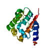
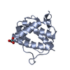
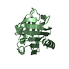
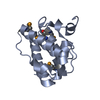
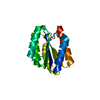
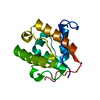
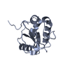
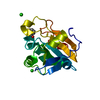
- PDB-1pa7: Crystal structure of amino-terminal microtubule binding domain of EB1 -
+ Open data

- Basic information
| Entry | Database: PDB / ID: 1pa7 | ||||||
|---|---|---|---|---|---|---|---|
| Title | Crystal structure of amino-terminal microtubule binding domain of EB1 | ||||||
 Components Components | Microtubule-associated protein RP/EB family member 1 | ||||||
 Keywords Keywords | STRUCTURAL PROTEIN / CH domain | ||||||
| Function / homology |  Function and homology information Function and homology informationprotein localization to astral microtubule / protein localization to mitotic spindle / cortical microtubule cytoskeleton / mitotic spindle astral microtubule end / protein localization to microtubule / cell projection membrane / microtubule plus-end / mitotic spindle microtubule / attachment of mitotic spindle microtubules to kinetochore / microtubule plus-end binding ...protein localization to astral microtubule / protein localization to mitotic spindle / cortical microtubule cytoskeleton / mitotic spindle astral microtubule end / protein localization to microtubule / cell projection membrane / microtubule plus-end / mitotic spindle microtubule / attachment of mitotic spindle microtubules to kinetochore / microtubule plus-end binding / microtubule bundle formation / non-motile cilium assembly / protein localization to centrosome / mitotic spindle pole / spindle midzone / establishment of mitotic spindle orientation / negative regulation of microtubule polymerization / microtubule polymerization / microtubule organizing center / regulation of microtubule polymerization or depolymerization / positive regulation of microtubule polymerization / cytoplasmic microtubule / spindle assembly / Amplification of signal from unattached kinetochores via a MAD2 inhibitory signal / Loss of Nlp from mitotic centrosomes / Loss of proteins required for interphase microtubule organization from the centrosome / Recruitment of mitotic centrosome proteins and complexes / Recruitment of NuMA to mitotic centrosomes / Anchoring of the basal body to the plasma membrane / Mitotic Prometaphase / EML4 and NUDC in mitotic spindle formation / AURKA Activation by TPX2 / Resolution of Sister Chromatid Cohesion / protein serine/threonine kinase binding / RHO GTPases Activate Formins / The role of GTSE1 in G2/M progression after G2 checkpoint / Separation of Sister Chromatids / intracellular protein localization / Regulation of PLK1 Activity at G2/M Transition / cell migration / microtubule / ciliary basal body / cadherin binding / cell division / focal adhesion / centrosome / Golgi apparatus / RNA binding / identical protein binding / cytosol Similarity search - Function | ||||||
| Biological species |  Homo sapiens (human) Homo sapiens (human) | ||||||
| Method |  X-RAY DIFFRACTION / SYNCHROTRON / MAD / Resolution: 1.45 Å X-RAY DIFFRACTION / SYNCHROTRON / MAD / Resolution: 1.45 Å | ||||||
 Authors Authors | Hayashi, I. / Ikura, M. | ||||||
 Citation Citation | Journal: J.Biol.Chem. / Year: 2003 Title: Crystal structure of the amino-terminal microtubule-binding domain of end-binding protein 1 (EB1) Authors: Hayashi, I. / Ikura, M. | ||||||
| History |
|
- Structure visualization
| Structure viewer | Molecule: MolmilJmol/JSmol |
|---|
- Downloads & links
-Download
| PDBx/mmCIF format | 1pa7.cif.gz | 41.4 KB | Display | PDBx/mmCIF format |
|---|---|---|---|---|
| PDB format | pdb1pa7.ent.gz | 28.9 KB | Display | PDB format |
| PDBx/mmJSON format | 1pa7.json.gz | Tree view | PDBx/mmJSON format | |
| Others |  Other downloads Other downloads |
-Validation report
| Arichive directory | https://data.pdbj.org/pub/pdb/validation_reports/pa/1pa7ftp://data.pdbj.org/pub/pdb/validation_reports/pa/1pa7 | HTTPS FTP |
|---|
-Related structure data
-Links
 PDBj
PDBj


- Assembly
| Deposited unit | 
| ||||||||
|---|---|---|---|---|---|---|---|---|---|
| 1 |
| ||||||||
| Unit cell |
|
-Components
| #1: Protein | Mass: 15050.383 Da / Num. of mol.: 1 / Fragment: N-terminal domain, EB1 microtubule-binding domain Source method: isolated from a genetically manipulated source Source: (gene. exp.) Homo sapiens (human) / Gene: EB1 (amino acids 1-130) / Plasmid: pET15b / Species (production host): Escherichia coli / Production host:  | ||
|---|---|---|---|
| #2: Chemical |   Mass: 96.063 Da / Num. of mol.: 2 / Source method: obtained synthetically / Formula: SO4 Mass: 96.063 Da / Num. of mol.: 2 / Source method: obtained synthetically / Formula: SO4#3: Water | ChemComp-HOH / |  Mass: 18.015 Da / Num. of mol.: 136 / Source method: isolated from a natural source / Formula: H2O Mass: 18.015 Da / Num. of mol.: 136 / Source method: isolated from a natural source / Formula: H2O |
-Experimental details
-Experiment
| Experiment | Method: X-RAY DIFFRACTION |
|---|
- Sample preparation
| Crystal | Density Matthews: 2 Å3/Da / Density % sol: 38.12 % | ||||||||||||||||||||||||||||||||||||||||||||||||||||||||
|---|---|---|---|---|---|---|---|---|---|---|---|---|---|---|---|---|---|---|---|---|---|---|---|---|---|---|---|---|---|---|---|---|---|---|---|---|---|---|---|---|---|---|---|---|---|---|---|---|---|---|---|---|---|---|---|---|---|
| Crystal grow | Temperature: 298 K / Method: vapor diffusion, sitting drop / pH: 6 Details: PEG4K, ammonium sulfate, MES, pH 6.0, VAPOR DIFFUSION, SITTING DROP, temperature 298K | ||||||||||||||||||||||||||||||||||||||||||||||||||||||||
| Crystal grow | *PLUS pH: 8 / Method: vapor diffusion, sitting drop | ||||||||||||||||||||||||||||||||||||||||||||||||||||||||
| Components of the solutions | *PLUS
|
-Data collection
| Diffraction | Mean temperature: 100 K |
|---|---|
| Diffraction source | Source: SYNCHROTRON / Site: NSLS  / Beamline: X8C / Wavelength: 0.98 Å / Beamline: X8C / Wavelength: 0.98 Å |
| Detector | Type: ADSC QUANTUM 4 / Detector: CCD / Date: Oct 7, 2002 / Details: mirror |
| Radiation | Protocol: MAD / Monochromatic (M) / Laue (L): M / Scattering type: x-ray |
| Radiation wavelength | Wavelength: 0.98 Å / Relative weight: 1 |
| Reflection | Resolution: 1.45→20 Å / Num. obs: 23798 / Rmerge(I) obs: 0.042 |
| Reflection shell | Resolution: 1.45→1.5 Å / Rmerge(I) obs: 0.228 / Num. unique all: 2278 |
| Reflection | *PLUS % possible obs: 99 % |
| Reflection shell | *PLUS % possible obs: 97.8 % / Rmerge(I) obs: 0.192 / Mean I/σ(I) obs: 6.99 |
- Processing
| Software |
| ||||||||||||||||||||||||||||||||||||||||||||||||||||||||||||||||||||||||||||||||||||||||||||||||||||
|---|---|---|---|---|---|---|---|---|---|---|---|---|---|---|---|---|---|---|---|---|---|---|---|---|---|---|---|---|---|---|---|---|---|---|---|---|---|---|---|---|---|---|---|---|---|---|---|---|---|---|---|---|---|---|---|---|---|---|---|---|---|---|---|---|---|---|---|---|---|---|---|---|---|---|---|---|---|---|---|---|---|---|---|---|---|---|---|---|---|---|---|---|---|---|---|---|---|---|---|---|---|
| Refinement | Method to determine structure: MAD / Resolution: 1.45→20 Å / Cor.coef. Fo:Fc: 0.957 / Cor.coef. Fo:Fc free: 0.946 / SU B: 0.932 / SU ML: 0.037 / TLS residual ADP flag: LIKELY RESIDUAL / Cross valid method: THROUGHOUT / σ(F): 0 / ESU R: 0.066 / ESU R Free: 0.064 / Stereochemistry target values: MAXIMUM LIKELIHOOD / Details: HYDROGENS HAVE BEEN ADDED IN THE RIDING POSITIONS
| ||||||||||||||||||||||||||||||||||||||||||||||||||||||||||||||||||||||||||||||||||||||||||||||||||||
| Solvent computation | Ion probe radii: 0.8 Å / Shrinkage radii: 0.8 Å / VDW probe radii: 1.4 Å / Solvent model: BABINET MODEL WITH MASK | ||||||||||||||||||||||||||||||||||||||||||||||||||||||||||||||||||||||||||||||||||||||||||||||||||||
| Displacement parameters | Biso mean: 12.199 Å2
| ||||||||||||||||||||||||||||||||||||||||||||||||||||||||||||||||||||||||||||||||||||||||||||||||||||
| Refinement step | Cycle: LAST / Resolution: 1.45→20 Å
| ||||||||||||||||||||||||||||||||||||||||||||||||||||||||||||||||||||||||||||||||||||||||||||||||||||
| Refine LS restraints |
| ||||||||||||||||||||||||||||||||||||||||||||||||||||||||||||||||||||||||||||||||||||||||||||||||||||
| LS refinement shell | Resolution: 1.448→1.486 Å / Total num. of bins used: 20 /
| ||||||||||||||||||||||||||||||||||||||||||||||||||||||||||||||||||||||||||||||||||||||||||||||||||||
| Refinement TLS params. | Method: refined / Origin x: 2.8131 Å / Origin y: 23.293 Å / Origin z: 32.6876 Å
| ||||||||||||||||||||||||||||||||||||||||||||||||||||||||||||||||||||||||||||||||||||||||||||||||||||
| Refinement | *PLUS Num. reflection obs: 21093 / % reflection Rfree: 5 % / Rfactor Rfree: 0.189 / Rfactor Rwork: 0.173 | ||||||||||||||||||||||||||||||||||||||||||||||||||||||||||||||||||||||||||||||||||||||||||||||||||||
| Solvent computation | *PLUS | ||||||||||||||||||||||||||||||||||||||||||||||||||||||||||||||||||||||||||||||||||||||||||||||||||||
| Displacement parameters | *PLUS | ||||||||||||||||||||||||||||||||||||||||||||||||||||||||||||||||||||||||||||||||||||||||||||||||||||
| Refine LS restraints | *PLUS
|
