 ムービー
ムービー コントローラー


+ データを開く

- 基本情報
基本情報
| 登録情報 | データベース: PDB / ID: 1pa7 | ||||||
|---|---|---|---|---|---|---|---|
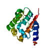
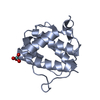
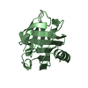
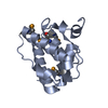
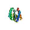
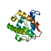
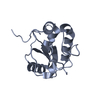
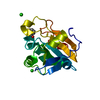
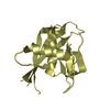
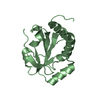
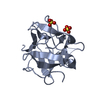
| タイトル | Crystal structure of amino-terminal microtubule binding domain of EB1 | ||||||
 要素 要素 | Microtubule-associated protein RP/EB family member 1 | ||||||
 キーワード キーワード |  STRUCTURAL PROTEIN (タンパク質) / CH domain STRUCTURAL PROTEIN (タンパク質) / CH domain | ||||||
| 機能・相同性 |  機能・相同性情報 機能・相同性情報protein localization to astral microtubule / cortical microtubule cytoskeleton / mitotic spindle astral microtubule end / protein localization to microtubule / microtubule plus-end / cell projection membrane / attachment of mitotic spindle microtubules to kinetochore / microtubule plus-end binding / non-motile cilium assembly / microtubule bundle formation ...protein localization to astral microtubule / cortical microtubule cytoskeleton / mitotic spindle astral microtubule end / protein localization to microtubule / microtubule plus-end / cell projection membrane / attachment of mitotic spindle microtubules to kinetochore / microtubule plus-end binding / non-motile cilium assembly / microtubule bundle formation / protein localization to centrosome / 微小管形成中心 / negative regulation of microtubule polymerization / mitotic spindle pole / 微小管 / establishment of mitotic spindle orientation / spindle assembly / regulation of microtubule polymerization or depolymerization / spindle midzone / cytoplasmic microtubule / Amplification of signal from unattached kinetochores via a MAD2 inhibitory signal / Mitotic Prometaphase / EML4 and NUDC in mitotic spindle formation / positive regulation of microtubule polymerization / Loss of Nlp from mitotic centrosomes / Loss of proteins required for interphase microtubule organization from the centrosome / Recruitment of mitotic centrosome proteins and complexes / Resolution of Sister Chromatid Cohesion / Recruitment of NuMA to mitotic centrosomes / Anchoring of the basal body to the plasma membrane / AURKA Activation by TPX2 / ciliary basal body / RHO GTPases Activate Formins / protein localization / Separation of Sister Chromatids / The role of GTSE1 in G2/M progression after G2 checkpoint / Regulation of PLK1 Activity at G2/M Transition / 遊走 / 微小管 / molecular adaptor activity / cadherin binding / 細胞分裂 / focal adhesion / 中心体 / protein kinase binding / ゴルジ体 / RNA binding / identical protein binding / 細胞質基質類似検索 - 分子機能 | ||||||
| 生物種 |  Homo sapiens (ヒト) Homo sapiens (ヒト) | ||||||
| 手法 | X線回折 / シンクロトロン / 多波長異常分散 / 解像度: 1.45 Å | ||||||
 データ登録者 データ登録者 | Hayashi, I. / Ikura, M. | ||||||
 引用 引用 | ジャーナル: J.Biol.Chem. / 年: 2003 タイトル: Crystal structure of the amino-terminal microtubule-binding domain of end-binding protein 1 (EB1) 著者: Hayashi, I. / Ikura, M. | ||||||
| 履歴 |
|
- 構造の表示
構造の表示
| 構造ビューア | 分子: MolmilJmol/JSmol |
|---|
- ダウンロードとリンク
-ダウンロード
| PDBx/mmCIF形式 | 1pa7.cif.gz | 41.4 KB | 表示 | PDBx/mmCIF形式 |
|---|---|---|---|---|
| PDB形式 | pdb1pa7.ent.gz | 28.9 KB | 表示 | PDB形式 |
| PDBx/mmJSON形式 | 1pa7.json.gz | ツリー表示 | PDBx/mmJSON形式 | |
| その他 |  その他のダウンロード その他のダウンロード |
-検証レポート
| アーカイブディレクトリ | https://data.pdbj.org/pub/pdb/validation_reports/pa/1pa7ftp://data.pdbj.org/pub/pdb/validation_reports/pa/1pa7 | HTTPS FTP |
|---|
-関連構造データ
-リンク
 PDBj
PDBj


- 集合体
集合体
| 登録構造単位 | 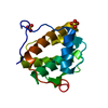
| ||||||||
|---|---|---|---|---|---|---|---|---|---|
| 1 |
| ||||||||
| 単位格子 |
|
-要素
| #1: タンパク質 | 分子量: 15050.383 Da / 分子数: 1 / 断片: N-terminal domain, EB1 microtubule-binding domain / 由来タイプ: 組換発現 / 由来: (組換発現) Homo sapiens (ヒト) / 遺伝子: EB1 (amino acids 1-130) / プラスミド: pET15b / 生物種 (発現宿主): Escherichia coli / 発現宿主:  Escherichia coli BL21(DE3) (大腸菌) / 株 (発現宿主): BL21(DE3) / 参照: UniProt: Q15691 Escherichia coli BL21(DE3) (大腸菌) / 株 (発現宿主): BL21(DE3) / 参照: UniProt: Q15691 | ||
|---|---|---|---|
| #2: 化合物 | 硫酸塩  分子量: 96.063 Da / 分子数: 2 / 由来タイプ: 合成 / 式: SO4 分子量: 96.063 Da / 分子数: 2 / 由来タイプ: 合成 / 式: SO4#3: 水 | ChemComp-HOH / | 水 分子量: 18.015 Da / 分子数: 136 / 由来タイプ: 天然 / 式: H2O 分子量: 18.015 Da / 分子数: 136 / 由来タイプ: 天然 / 式: H2O |
-実験情報
-実験
| 実験 | 手法: X線回折 |
|---|
- 試料調製
試料調製
| 結晶 | マシュー密度: 2 Å3/Da / 溶媒含有率: 38.12 % | ||||||||||||||||||||||||||||||||||||||||||||||||||||||||
|---|---|---|---|---|---|---|---|---|---|---|---|---|---|---|---|---|---|---|---|---|---|---|---|---|---|---|---|---|---|---|---|---|---|---|---|---|---|---|---|---|---|---|---|---|---|---|---|---|---|---|---|---|---|---|---|---|---|
| 結晶化 | 温度: 298 K / 手法: 蒸気拡散法, シッティングドロップ法 / pH: 6 詳細: PEG4K, ammonium sulfate, MES, pH 6.0, VAPOR DIFFUSION, SITTING DROP, temperature 298K | ||||||||||||||||||||||||||||||||||||||||||||||||||||||||
| 結晶化 | *PLUS pH: 8 / 手法: 蒸気拡散法, シッティングドロップ法 | ||||||||||||||||||||||||||||||||||||||||||||||||||||||||
| 溶液の組成 | *PLUS
|
-データ収集
| 回折 | 平均測定温度: 100 K |
|---|---|
| 放射光源 | 由来: シンクロトロン / サイト: NSLS  / ビームライン: X8C / 波長: 0.98 Å / ビームライン: X8C / 波長: 0.98 Å |
| 検出器 | タイプ: ADSC QUANTUM 4 / 検出器: CCD / 日付: 2002年10月7日 / 詳細: mirror |
| 放射 | プロトコル: MAD / 単色(M)・ラウエ(L): M / 散乱光タイプ: x-ray |
| 放射波長 | 波長: 0.98 Å / 相対比: 1 |
| 反射 | 解像度: 1.45→20 Å / Num. obs: 23798 / Rmerge(I) obs: 0.042 |
| 反射 シェル | 解像度: 1.45→1.5 Å / Rmerge(I) obs: 0.228 / Num. unique all: 2278 |
| 反射 | *PLUS % possible obs: 99 % |
| 反射 シェル | *PLUS % possible obs: 97.8 % / Rmerge(I) obs: 0.192 / Mean I/σ(I) obs: 6.99 |
- 解析
解析
| ソフトウェア |
| ||||||||||||||||||||||||||||||||||||||||||||||||||||||||||||||||||||||||||||||||||||||||||||||||||||
|---|---|---|---|---|---|---|---|---|---|---|---|---|---|---|---|---|---|---|---|---|---|---|---|---|---|---|---|---|---|---|---|---|---|---|---|---|---|---|---|---|---|---|---|---|---|---|---|---|---|---|---|---|---|---|---|---|---|---|---|---|---|---|---|---|---|---|---|---|---|---|---|---|---|---|---|---|---|---|---|---|---|---|---|---|---|---|---|---|---|---|---|---|---|---|---|---|---|---|---|---|---|
| 精密化 | 構造決定の手法: 多波長異常分散 / 解像度: 1.45→20 Å / Cor.coef. Fo:Fc: 0.957 / Cor.coef. Fo:Fc free: 0.946 / SU B: 0.932 / SU ML: 0.037 / TLS residual ADP flag: LIKELY RESIDUAL / 交差検証法: THROUGHOUT / σ(F): 0 / ESU R: 0.066 / ESU R Free: 0.064 / 立体化学のターゲット値: MAXIMUM LIKELIHOOD / 詳細: HYDROGENS HAVE BEEN ADDED IN THE RIDING POSITIONS
| ||||||||||||||||||||||||||||||||||||||||||||||||||||||||||||||||||||||||||||||||||||||||||||||||||||
| 溶媒の処理 | イオンプローブ半径: 0.8 Å / 減衰半径: 0.8 Å / VDWプローブ半径: 1.4 Å / 溶媒モデル: BABINET MODEL WITH MASK | ||||||||||||||||||||||||||||||||||||||||||||||||||||||||||||||||||||||||||||||||||||||||||||||||||||
| 原子変位パラメータ | Biso mean: 12.199 Å2
| ||||||||||||||||||||||||||||||||||||||||||||||||||||||||||||||||||||||||||||||||||||||||||||||||||||
| 精密化ステップ | サイクル: LAST / 解像度: 1.45→20 Å
| ||||||||||||||||||||||||||||||||||||||||||||||||||||||||||||||||||||||||||||||||||||||||||||||||||||
| 拘束条件 |
| ||||||||||||||||||||||||||||||||||||||||||||||||||||||||||||||||||||||||||||||||||||||||||||||||||||
| LS精密化 シェル | 解像度: 1.448→1.486 Å / Total num. of bins used: 20 /
| ||||||||||||||||||||||||||||||||||||||||||||||||||||||||||||||||||||||||||||||||||||||||||||||||||||
| 精密化 TLS | 手法: refined / Origin x: 2.8131 Å / Origin y: 23.293 Å / Origin z: 32.6876 Å
| ||||||||||||||||||||||||||||||||||||||||||||||||||||||||||||||||||||||||||||||||||||||||||||||||||||
| 精密化 | *PLUS Num. reflection obs: 21093 / % reflection Rfree: 5 % / Rfactor Rfree: 0.189 / Rfactor Rwork: 0.173 | ||||||||||||||||||||||||||||||||||||||||||||||||||||||||||||||||||||||||||||||||||||||||||||||||||||
| 溶媒の処理 | *PLUS | ||||||||||||||||||||||||||||||||||||||||||||||||||||||||||||||||||||||||||||||||||||||||||||||||||||
| 原子変位パラメータ | *PLUS | ||||||||||||||||||||||||||||||||||||||||||||||||||||||||||||||||||||||||||||||||||||||||||||||||||||
| 拘束条件 | *PLUS
|
