 Movie
Movie Controller


+ Open data

- Basic information
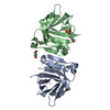
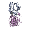
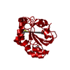
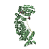
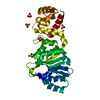
| Entry | Database: PDB / ID: 3kza | ||||||
|---|---|---|---|---|---|---|---|
| Title | Crystal structure of Gyuba, a patched chimera of b-lactglobulin | ||||||
 Components Components | Beta-lactoglobulin | ||||||
 Keywords Keywords | TRANSPORT PROTEIN / artificial protein / chimera protein / Disulfide bond / Milk protein / Retinol-binding / Secreted / Transport | ||||||
| Function / homology |  Function and homology information Function and homology informationretinol binding / long-chain fatty acid binding / extracellular region / identical protein binding Similarity search - Function | ||||||
| Biological species |  | ||||||
| Method |  X-RAY DIFFRACTION / SYNCHROTRON / MOLECULAR REPLACEMENT / Resolution: 2 Å X-RAY DIFFRACTION / SYNCHROTRON / MOLECULAR REPLACEMENT / Resolution: 2 Å | ||||||
 Authors Authors | Tsuge, H. / Ohtomo, H. / Utsunomiya, H. / Konuma, T. / Ikeguchi, M. | ||||||
 Citation Citation | Journal: Protein Sci. / Year: 2011 Title: Structure and stability of Gyuba, a patched chimera of b-lactoglobulin Authors: Ohtomo, H. / Konuma, T. / Utsunoiya, H. / Tsuge, H. / Ikeguchi, M. | ||||||
| History |
|
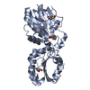
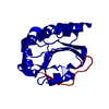
- Structure visualization
| Structure viewer | Molecule: MolmilJmol/JSmol |
|---|
- Downloads & links
-Download
| PDBx/mmCIF format | 3kza.cif.gz | 76.5 KB | Display | PDBx/mmCIF format |
|---|---|---|---|---|
| PDB format | pdb3kza.ent.gz | 57.6 KB | Display | PDB format |
| PDBx/mmJSON format | 3kza.json.gz | Tree view | PDBx/mmJSON format | |
| Others |  Other downloads Other downloads |
-Validation report
| Arichive directory | https://data.pdbj.org/pub/pdb/validation_reports/kz/3kzaftp://data.pdbj.org/pub/pdb/validation_reports/kz/3kza | HTTPS FTP |
|---|
-Related structure data
| Similar structure data |
|---|


-Links
 PDBj
PDBj

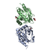
- Assembly
| Deposited unit | 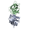
| ||||||||
|---|---|---|---|---|---|---|---|---|---|
| 1 |
| ||||||||
| 2 | 
| ||||||||
| 3 | 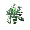
| ||||||||
| Unit cell |
|
-Components
| #1: Protein | Mass: 18310.939 Da / Num. of mol.: 2 Source method: isolated from a genetically manipulated source Details: AN ARTIFICIAL CHIMERA PROTEIN OF BOS-TAURAS AND EQUINE B-LACTOGLOBULIN. THE RESIDUES 1-16, 28-41, 50, 51, 63-65, 74-49, 87, 88, 96-101, 109-116, 125-129, 139-144, 154-162 ARE FROM EQUINE, ...Details: AN ARTIFICIAL CHIMERA PROTEIN OF BOS-TAURAS AND EQUINE B-LACTOGLOBULIN. THE RESIDUES 1-16, 28-41, 50, 51, 63-65, 74-49, 87, 88, 96-101, 109-116, 125-129, 139-144, 154-162 ARE FROM EQUINE, THE RESIDUES 17-27, 42-49, 52-62, 66-73, 80-86, 89-95, 102-108, 117-124, 130-138, 145-153 ARE FROM BOS-TAURAS. Source: (gene. exp.) Gene: LGB1 / Plasmid: pET3c / Production host:  #2: Water | ChemComp-HOH / |  Mass: 18.015 Da / Num. of mol.: 240 / Source method: isolated from a natural source / Formula: H2O Mass: 18.015 Da / Num. of mol.: 240 / Source method: isolated from a natural source / Formula: H2OHas protein modification | Y | Sequence details | THIS IS AN ARTIFICIAL | |
|---|
-Experimental details
-Experiment
| Experiment | Method: X-RAY DIFFRACTION / Number of used crystals: 1 |
|---|
- Sample preparation
| Crystal | Density Matthews: 2.84 Å3/Da / Density % sol: 56.67 % |
|---|---|
| Crystal grow | Temperature: 293 K / Method: vapor diffusion, hanging drop / pH: 7 Details: 2.0M ammonium sulfate, pH 7.0, VAPOR DIFFUSION, HANGING DROP, temperature 293K |
-Data collection
| Diffraction | Mean temperature: 277 K |
|---|---|
| Diffraction source | Source: SYNCHROTRON / Site: Photon Factory  / Beamline: AR-NW12A / Beamline: AR-NW12A |
| Detector | Type: ADSC QUANTUM 210 / Detector: CCD |
| Radiation | Protocol: SINGLE WAVELENGTH / Monochromatic (M) / Laue (L): M / Scattering type: x-ray |
| Radiation wavelength | Relative weight: 1 |
| Reflection | Resolution: 2→59.66 Å / Num. obs: 27566 / % possible obs: 99.8 % / Observed criterion σ(F): 1 / Observed criterion σ(I): 1 / Redundancy: 10.9 % / Rsym value: 0.066 / Net I/σ(I): 15.2 |
| Reflection shell | Resolution: 2→2.07 Å / Redundancy: 9.6 % / Num. unique all: 2741 / Rsym value: 0.314 / % possible all: 99.9 |
- Processing
| Software |
| ||||||||||||||||||||||||||||||||||||||||||||||||||||||||||||||||||||||||||||||||||||||||||
|---|---|---|---|---|---|---|---|---|---|---|---|---|---|---|---|---|---|---|---|---|---|---|---|---|---|---|---|---|---|---|---|---|---|---|---|---|---|---|---|---|---|---|---|---|---|---|---|---|---|---|---|---|---|---|---|---|---|---|---|---|---|---|---|---|---|---|---|---|---|---|---|---|---|---|---|---|---|---|---|---|---|---|---|---|---|---|---|---|---|---|---|
| Refinement | Method to determine structure: MOLECULAR REPLACEMENT / Resolution: 2→59.66 Å / Cor.coef. Fo:Fc: 0.935 / Cor.coef. Fo:Fc free: 0.91 / SU B: 4.16 / SU ML: 0.121 / Cross valid method: THROUGHOUT / ESU R: 0.193 / ESU R Free: 0.182 / Stereochemistry target values: MAXIMUM LIKELIHOOD / Details: HYDROGENS HAVE BEEN ADDED IN THE RIDING POSITIONS
| ||||||||||||||||||||||||||||||||||||||||||||||||||||||||||||||||||||||||||||||||||||||||||
| Solvent computation | Ion probe radii: 0.8 Å / Shrinkage radii: 0.8 Å / VDW probe radii: 1.4 Å / Solvent model: MASK | ||||||||||||||||||||||||||||||||||||||||||||||||||||||||||||||||||||||||||||||||||||||||||
| Displacement parameters | Biso mean: 35.665 Å2
| ||||||||||||||||||||||||||||||||||||||||||||||||||||||||||||||||||||||||||||||||||||||||||
| Refinement step | Cycle: LAST / Resolution: 2→59.66 Å
| ||||||||||||||||||||||||||||||||||||||||||||||||||||||||||||||||||||||||||||||||||||||||||
| Refine LS restraints |
| ||||||||||||||||||||||||||||||||||||||||||||||||||||||||||||||||||||||||||||||||||||||||||
| LS refinement shell | Resolution: 1.998→2.05 Å / Total num. of bins used: 20
|
