 Movie
Movie Controller

[English] 日本語
 Yorodumi
- PDB-1p9l: Structure of M. tuberculosis dihydrodipicolinate reductase in com... -
+ Open data

- Basic information
| Entry | Database: PDB / ID: 1p9l | ||||||
|---|---|---|---|---|---|---|---|
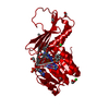
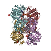
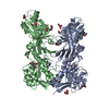
| Title | Structure of M. tuberculosis dihydrodipicolinate reductase in complex with NADH and 2,6 PDC | ||||||
 Components Components | dihydrodipicolinate reductase | ||||||
 Keywords Keywords | OXIDOREDUCTASE / REDUCTASE / LYSINE BIOSYNTHESIS / NADH BINDING SPECIFICITY / TB Structural Genomics Consortium / TBSGC / Structural Genomics / PSI / Protein Structure Initiative | ||||||
| Function / homology |  Function and homology information Function and homology information4-hydroxy-tetrahydrodipicolinate reductase / oxidoreductase activity, acting on CH or CH2 groups, NAD or NADP as acceptor / 4-hydroxy-tetrahydrodipicolinate reductase activity / cell wall / NADH binding / : / : / NADPH binding / peptidoglycan-based cell wall / plasma membrane / cytosol Similarity search - Function | ||||||
| Biological species |   Mycobacterium tuberculosis (bacteria) Mycobacterium tuberculosis (bacteria) | ||||||
| Method |  X-RAY DIFFRACTION / MOLECULAR REPLACEMENT / Resolution: 2.3 Å X-RAY DIFFRACTION / MOLECULAR REPLACEMENT / Resolution: 2.3 Å | ||||||
 Authors Authors | Cirilli, M. / Zheng, R. / Scapin, G. / Blanchard, J.S. / TB Structural Genomics Consortium (TBSGC) | ||||||
 Citation Citation | Journal: Biochemistry / Year: 2003 Title: The three-dimensional structures of the Mycobacterium tuberculosis dihydrodipicolinate reductase-NADH-2,6-PDC and -NADPH-2,6-PDC complexes. Structural and mutagenic analysis of relaxed nucleotide specificity. Authors: Cirilli, M. / Zheng, R. / Scapin, G. / Blanchard, J.S. | ||||||
| History |
|
- Structure visualization
| Structure viewer | Molecule: MolmilJmol/JSmol |
|---|
- Downloads & links
-Download
| PDBx/mmCIF format | 1p9l.cif.gz | 107.3 KB | Display | PDBx/mmCIF format |
|---|---|---|---|---|
| PDB format | pdb1p9l.ent.gz | 83 KB | Display | PDB format |
| PDBx/mmJSON format | 1p9l.json.gz | Tree view | PDBx/mmJSON format | |
| Others |  Other downloads Other downloads |
-Validation report
| Arichive directory | https://data.pdbj.org/pub/pdb/validation_reports/p9/1p9lftp://data.pdbj.org/pub/pdb/validation_reports/p9/1p9l | HTTPS FTP |
|---|
-Related structure data
| Related structure data |  1c3vC 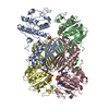 1arzS S: Starting model for refinement C: citing same article ( |
|---|---|
| Similar structure data | |
| Other databases |


-Links
 PDBj
PDBj

- Assembly
| Deposited unit | 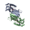
| ||||||||
|---|---|---|---|---|---|---|---|---|---|
| 1 | 
| ||||||||
| Unit cell |
| ||||||||
| Details | The biological assembly is a tetramer, generated by a crystallographic two-fold axis |
-Components
| #1: Protein | Mass: 25793.383 Da / Num. of mol.: 2 Source method: isolated from a genetically manipulated source Source: (gene. exp.) Mycobacterium tuberculosis (bacteria) / Gene: dapB / Plasmid: pET23a(+) / Species (production host): Escherichia coli / Production host: References: UniProt: P72024, UniProt: P9WP23*PLUS, EC: 1.3.1.26 #2: Chemical |   Mass: 665.441 Da / Num. of mol.: 2 / Source method: obtained synthetically / Formula: C21H29N7O14P2 Mass: 665.441 Da / Num. of mol.: 2 / Source method: obtained synthetically / Formula: C21H29N7O14P2#3: Chemical |   Mass: 167.119 Da / Num. of mol.: 2 / Source method: obtained synthetically / Formula: C7H5NO4 Mass: 167.119 Da / Num. of mol.: 2 / Source method: obtained synthetically / Formula: C7H5NO4#4: Chemical | ChemComp-PG4 / |   Mass: 194.226 Da / Num. of mol.: 1 / Source method: obtained synthetically / Formula: C8H18O5 / Comment: precipitant*YM Mass: 194.226 Da / Num. of mol.: 1 / Source method: obtained synthetically / Formula: C8H18O5 / Comment: precipitant*YM#5: Water | ChemComp-HOH / |  Mass: 18.015 Da / Num. of mol.: 117 / Source method: isolated from a natural source / Formula: H2O Mass: 18.015 Da / Num. of mol.: 117 / Source method: isolated from a natural source / Formula: H2O |
|---|
-Experimental details
-Experiment
| Experiment | Method: X-RAY DIFFRACTION / Number of used crystals: 1 |
|---|
- Sample preparation
| Crystal | Density Matthews: 2.66 Å3/Da / Density % sol: 53.78 % | ||||||||||||||||||||||||||||||||||||||||||
|---|---|---|---|---|---|---|---|---|---|---|---|---|---|---|---|---|---|---|---|---|---|---|---|---|---|---|---|---|---|---|---|---|---|---|---|---|---|---|---|---|---|---|---|
| Crystal grow | Temperature: 298 K / Method: vapor diffusion, hanging drop / pH: 7.5 Details: ammonium Sulfate, PEG 400, Hepes, pH 7.5, VAPOR DIFFUSION, HANGING DROP, temperature 298K | ||||||||||||||||||||||||||||||||||||||||||
| Crystal grow | *PLUS Method: vapor diffusion, hanging drop | ||||||||||||||||||||||||||||||||||||||||||
| Components of the solutions | *PLUS
|
-Data collection
| Diffraction | Mean temperature: 298 K |
|---|---|
| Diffraction source | Source: ROTATING ANODE / Type: RIGAKU RU200 / Wavelength: 1.5418 Å |
| Detector | Type: SIEMENS / Detector: AREA DETECTOR / Date: Sep 12, 1996 |
| Radiation | Monochromator: Ni filter / Protocol: SINGLE WAVELENGTH / Monochromatic (M) / Laue (L): M / Scattering type: x-ray |
| Radiation wavelength | Wavelength: 1.5418 Å / Relative weight: 1 |
| Reflection | Resolution: 2.3→25 Å / Num. all: 24785 / Num. obs: 24502 / % possible obs: 98.5 % / Observed criterion σ(F): 0 / Observed criterion σ(I): -3 / Redundancy: 3.2 % / Biso Wilson estimate: 28.7 Å2 / Rsym value: 0.078 / Net I/σ(I): 13.6 |
| Reflection shell | Resolution: 2.3→2.4 Å / Redundancy: 3.2 % / Mean I/σ(I) obs: 3.3 / Num. unique all: 2353 / Rsym value: 0.206 / % possible all: 95.1 |
| Reflection | *PLUS Lowest resolution: 25 Å / Num. measured all: 77959 / Rmerge(I) obs: 0.078 |
| Reflection shell | *PLUS % possible obs: 95.1 % / Redundancy: 2.1 % / Num. unique obs: 2353 / Num. measured obs: 4910 / Rmerge(I) obs: 0.206 |
- Processing
| Software |
| ||||||||||||||||||||||||||||||||||||
|---|---|---|---|---|---|---|---|---|---|---|---|---|---|---|---|---|---|---|---|---|---|---|---|---|---|---|---|---|---|---|---|---|---|---|---|---|---|
| Refinement | Method to determine structure: MOLECULAR REPLACEMENT Starting model: 1ARZ Resolution: 2.3→25 Å / Data cutoff high absF: 0.001 / Data cutoff low absF: 10000000 / Isotropic thermal model: restrained / Cross valid method: THROUGHOUT / σ(F): 1 / Stereochemistry target values: Engh & Huber
| ||||||||||||||||||||||||||||||||||||
| Displacement parameters | Biso mean: 26.8 Å2 | ||||||||||||||||||||||||||||||||||||
| Refinement step | Cycle: LAST / Resolution: 2.3→25 Å
| ||||||||||||||||||||||||||||||||||||
| Refine LS restraints |
| ||||||||||||||||||||||||||||||||||||
| LS refinement shell | Resolution: 2.3→2.4 Å
| ||||||||||||||||||||||||||||||||||||
| Refinement | *PLUS Rfactor Rfree: 0.25 | ||||||||||||||||||||||||||||||||||||
| Solvent computation | *PLUS | ||||||||||||||||||||||||||||||||||||
| Displacement parameters | *PLUS | ||||||||||||||||||||||||||||||||||||
| Refine LS restraints | *PLUS
|
