 Movie
Movie Controller

[English] 日本語
 Yorodumi
- PDB-1p3h: Crystal Structure of the Mycobacterium tuberculosis chaperonin 10... -
+ Open data

- Basic information
| Entry | Database: PDB / ID: 1p3h | |||||||||
|---|---|---|---|---|---|---|---|---|---|---|
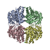
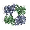
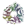
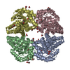
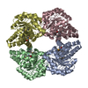
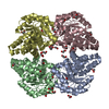
| Title | Crystal Structure of the Mycobacterium tuberculosis chaperonin 10 tetradecamer | |||||||||
 Components Components | 10 kDa chaperonin | |||||||||
 Keywords Keywords | CHAPERONE / BETA BARREL / ACIDIC CLUSTER / FLEXIBLE LOOP / Structural Genomics / PSI / Protein Structure Initiative / TB Structural Genomics Consortium / TBSGC | |||||||||
| Function / homology |  Function and homology information Function and homology information: / cell wall / zymogen binding / protein folding chaperone / peptidoglycan-based cell wall / : / cellular response to heat / response to heat / protein-folding chaperone binding / response to antibiotic ...: / cell wall / zymogen binding / protein folding chaperone / peptidoglycan-based cell wall / : / cellular response to heat / response to heat / protein-folding chaperone binding / response to antibiotic / regulation of DNA-templated transcription / ATP hydrolysis activity / extracellular region / ATP binding / metal ion binding / plasma membrane / cytosol Similarity search - Function | |||||||||
| Biological species |   Mycobacterium tuberculosis (bacteria) Mycobacterium tuberculosis (bacteria) | |||||||||
| Method |  X-RAY DIFFRACTION / SYNCHROTRON / MOLECULAR REPLACEMENT / Resolution: 2.8 Å X-RAY DIFFRACTION / SYNCHROTRON / MOLECULAR REPLACEMENT / Resolution: 2.8 Å | |||||||||
 Authors Authors | Roberts, M.M. / Coker, A.R. / Fossati, G. / Mascagni, P. / Coates, A.R.M. / Wood, S.P. / TB Structural Genomics Consortium (TBSGC) | |||||||||
 Citation Citation | Journal: J.BACTERIOL. / Year: 2003 Title: Mycobacterium tuberculosis chaperonin 10 heptamers self-associate through their biologically active loops Authors: Roberts, M.M. / Coker, A.R. / Fossati, G. / Mascagni, P. / Coates, A.R.M. / Wood, S.P. #1: Journal: Acta Crystallogr.,Sect.D / Year: 1999Title: Crystallization, X-ray diffraction and preliminary structure analysis of Mycobacterium tuberculosis chaperonin 10 Authors: Roberts, M.M. / Coker, A.R. / Fossati, G. / Mascagni, P. / Coates, A.R.M. / Wood, S.P. #2: Journal: J.EXP.MED. / Year: 1997Title: Mycobacterium tuberculosis chaperonin 10 stimulates bone resorption: A potential contributory factor in Pott's Disease Authors: Meghji, S. / White, P.A. / Nair, S.P. / Reddi, K. / Heron, K. / Henderson, B. / Zaliani, A. / Fossati, G. / Mascagni, P. / Hunt, J.F. / Roberts, M.M. / Coates, A.R.M. | |||||||||
| History |
|
- Structure visualization
| Structure viewer | Molecule: MolmilJmol/JSmol |
|---|
- Downloads & links
-Download
| PDBx/mmCIF format | 1p3h.cif.gz | 268.7 KB | Display | PDBx/mmCIF format |
|---|---|---|---|---|
| PDB format | pdb1p3h.ent.gz | 221.4 KB | Display | PDB format |
| PDBx/mmJSON format | 1p3h.json.gz | Tree view | PDBx/mmJSON format | |
| Others |  Other downloads Other downloads |
-Validation report
| Arichive directory | https://data.pdbj.org/pub/pdb/validation_reports/p3/1p3hftp://data.pdbj.org/pub/pdb/validation_reports/p3/1p3h | HTTPS FTP |
|---|
-Related structure data
| Similar structure data | |
|---|---|
| Other databases |


-Links
 PDBj
PDBj

- Assembly
| Deposited unit | 
| |||||||||
|---|---|---|---|---|---|---|---|---|---|---|
| 1 | 
| |||||||||
| 2 | 
| |||||||||
| 3 |
| |||||||||
| Unit cell |
| |||||||||
| Noncrystallographic symmetry (NCS) | NCS domain:
|
-Components
| #1: Protein | Mass: 10684.979 Da / Num. of mol.: 14 Source method: isolated from a genetically manipulated source Source: (gene. exp.) Mycobacterium tuberculosis (bacteria)Gene: GROS OR GROES OR MOPB OR CPN10 OR RV3418C OR MT3527 OR MTCY78.11 Plasmid: pUC18 / Species (production host): Escherichia coli / Production host: #2: Chemical | ChemComp-CA / |   Mass: 40.078 Da / Num. of mol.: 1 / Source method: obtained synthetically / Formula: Ca Mass: 40.078 Da / Num. of mol.: 1 / Source method: obtained synthetically / Formula: Ca#3: Chemical | ChemComp-MPD / (   Mass: 118.174 Da / Num. of mol.: 18 / Source method: obtained synthetically / Formula: C6H14O2 / Comment: precipitant*YM Mass: 118.174 Da / Num. of mol.: 18 / Source method: obtained synthetically / Formula: C6H14O2 / Comment: precipitant*YM#4: Water | ChemComp-HOH / |  Mass: 18.015 Da / Num. of mol.: 64 / Source method: isolated from a natural source / Formula: H2O Mass: 18.015 Da / Num. of mol.: 64 / Source method: isolated from a natural source / Formula: H2O |
|---|
-Experimental details
-Experiment
| Experiment | Method: X-RAY DIFFRACTION / Number of used crystals: 1 |
|---|
- Sample preparation
| Crystal | Density Matthews: 2.67 Å3/Da / Density % sol: 53.66 % | |||||||||||||||||||||||||||||||||||
|---|---|---|---|---|---|---|---|---|---|---|---|---|---|---|---|---|---|---|---|---|---|---|---|---|---|---|---|---|---|---|---|---|---|---|---|---|
| Crystal grow | Temperature: 294 K / Method: vapor diffusion, sitting drop / pH: 5.4 Details: MPD, sodium acetate, calcium chloride, pH 5.4, VAPOR DIFFUSION, SITTING DROP, temperature 294K | |||||||||||||||||||||||||||||||||||
| Crystal grow | *PLUS Method: vapor diffusion, sitting dropDetails: Roberts, M.M., (1999) Acta Crystallogr.,Sect.D, 55, 910. | |||||||||||||||||||||||||||||||||||
| Components of the solutions | *PLUS
|
-Data collection
| Diffraction | Mean temperature: 100 K |
|---|---|
| Diffraction source | Source: SYNCHROTRON / Site: SRS  / Beamline: PX7.2 / Wavelength: 1.488 Å / Beamline: PX7.2 / Wavelength: 1.488 Å |
| Detector | Type: MARRESEARCH / Detector: IMAGE PLATE / Date: Apr 8, 1996 / Details: mirrors |
| Radiation | Monochromator: GERMANIUM CRYSTAL SET TO BRAGG REFLECTION (111) Protocol: SINGLE WAVELENGTH / Monochromatic (M) / Laue (L): M / Scattering type: x-ray |
| Radiation wavelength | Wavelength: 1.488 Å / Relative weight: 1 |
| Reflection | Resolution: 2.8→30 Å / Num. all: 38321 / Num. obs: 38321 / % possible obs: 98 % / Observed criterion σ(F): 0 / Observed criterion σ(I): 0 / Redundancy: 3.5 % / Biso Wilson estimate: 80.3 Å2 / Rmerge(I) obs: 0.05 / Rsym value: 0.05 / Net I/σ(I): 24.41 |
| Reflection shell | Resolution: 2.8→2.82 Å / Redundancy: 3.5 % / Rmerge(I) obs: 0.61 / Mean I/σ(I) obs: 1.92 / Num. unique all: 982 / Rsym value: 0.61 / % possible all: 99.7 |
| Reflection | *PLUS Num. measured all: 134674 |
| Reflection shell | *PLUS % possible obs: 99.7 % / Rmerge(I) obs: 0.611 / Mean I/σ(I) obs: 1.6 |
- Processing
| Software |
| ||||||||||||||||||||||||||||||||||||
|---|---|---|---|---|---|---|---|---|---|---|---|---|---|---|---|---|---|---|---|---|---|---|---|---|---|---|---|---|---|---|---|---|---|---|---|---|---|
| Refinement | Method to determine structure: MOLECULAR REPLACEMENT Starting model: THE E.COLI GROES STRUCTURE WITH AMINO ACIDS DIFFERING FROM M.TUBERCULOSIS CHAPERONIN 10 TRUNCATED TO ALANINES Resolution: 2.8→30 Å / Rfactor Rfree error: 0.006 / Data cutoff high absF: 10000 / Data cutoff high rms absF: 10000 / Isotropic thermal model: restrained Cross valid method: TEST SET OF REFLECTIONS NOT USED IN REFINEMENT BUT TO MONITOR R-FREE σ(F): 0 / σ(I): 0 / Stereochemistry target values: Engh & Huber Details: RMS VALUES FOR NCS RELATED SUBUNITS APPLY TO ALL ATOMS IN EACH SUBUNIT. THE POSITIONAL NCS WEIGHTS INDICATED APPLY RESPECTIVELY TO MAINCHAIN (group 1) AND SIDECHAIN (group 2) ATOMS.
| ||||||||||||||||||||||||||||||||||||
| Solvent computation | Solvent model: flat model / Bsol: 51.7 Å2 / ksol: 0.263 e/Å3 | ||||||||||||||||||||||||||||||||||||
| Displacement parameters | Biso mean: 75.8 Å2
| ||||||||||||||||||||||||||||||||||||
| Refine analyze |
| ||||||||||||||||||||||||||||||||||||
| Refinement step | Cycle: LAST / Resolution: 2.8→30 Å
| ||||||||||||||||||||||||||||||||||||
| Refine LS restraints |
| ||||||||||||||||||||||||||||||||||||
| Refine LS restraints NCS | Refine-ID: X-RAY DIFFRACTION / Rms dev Biso : 13.85 Å2 / Rms dev position: 1.33 Å / Weight Biso : 1
| ||||||||||||||||||||||||||||||||||||
| LS refinement shell | Resolution: 2.8→2.98 Å / Rfactor Rfree error: 0.022 / Total num. of bins used: 6
| ||||||||||||||||||||||||||||||||||||
| Xplor file |
| ||||||||||||||||||||||||||||||||||||
| Refinement | *PLUS Rfactor Rfree: 0.28 | ||||||||||||||||||||||||||||||||||||
| Solvent computation | *PLUS | ||||||||||||||||||||||||||||||||||||
| Displacement parameters | *PLUS | ||||||||||||||||||||||||||||||||||||
| Refine LS restraints | *PLUS
|
