 Movie
Movie Controller

[English] 日本語
 Yorodumi
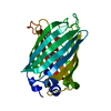
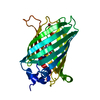
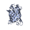
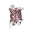
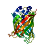
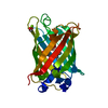
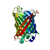
- PDB-1oxd: Expansion of the Genetic Code Enables Design of a Novel "Gold" Cl... -
+ Open data

- Basic information
| Entry | Database: PDB / ID: 1oxd | |||||||||
|---|---|---|---|---|---|---|---|---|---|---|
| Title | Expansion of the Genetic Code Enables Design of a Novel "Gold" Class of Green Fluorescent Proteins | |||||||||
 Components Components | cyan fluorescent protein cfp | |||||||||
 Keywords Keywords | LUMINESCENT PROTEIN / green fluorescent protein / chromophore / amino acid incorporation / tryptophan / genetic code | |||||||||
| Function / homology | Green Fluorescent Protein / Green fluorescent protein / Beta Barrel / Mainly Beta / :  Function and homology information Function and homology information | |||||||||
| Biological species | cfp marker plasmid pWM1009 (others) | |||||||||
| Method |  X-RAY DIFFRACTION / SYNCHROTRON / MOLECULAR REPLACEMENT / Resolution: 1.15 Å X-RAY DIFFRACTION / SYNCHROTRON / MOLECULAR REPLACEMENT / Resolution: 1.15 Å | |||||||||
 Authors Authors | Hyun Bae, J. / Rubini, M. / Jung, G. / Wiegand, G. / Seifert, M.H. / Azim, M.K. / Kim, J.S. / Zumbusch, A. / Holak, T.A. / Moroder, L. ...Hyun Bae, J. / Rubini, M. / Jung, G. / Wiegand, G. / Seifert, M.H. / Azim, M.K. / Kim, J.S. / Zumbusch, A. / Holak, T.A. / Moroder, L. / Huber, R. / Budisa, N. | |||||||||
 Citation Citation | Journal: J.Mol.Biol. / Year: 2003 Title: Expansion of the Genetic Code Enables Design of a Novel "Gold" Class of Green Fluorescent Proteins Authors: Hyun Bae, J. / Rubini, M. / Jung, G. / Wiegand, G. / Seifert, M.H. / Azim, M.K. / Kim, J.S. / Zumbusch, A. / Holak, T.A. / Moroder, L. / Huber, R. / Budisa, N. | |||||||||
| History |
|
- Structure visualization
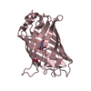
| Structure viewer | Molecule: MolmilJmol/JSmol |
|---|
- Downloads & links
-Download
| PDBx/mmCIF format | 1oxd.cif.gz | 60.7 KB | Display | PDBx/mmCIF format |
|---|---|---|---|---|
| PDB format | pdb1oxd.ent.gz | 43.4 KB | Display | PDB format |
| PDBx/mmJSON format | 1oxd.json.gz | Tree view | PDBx/mmJSON format | |
| Others |  Other downloads Other downloads |
-Validation report
| Arichive directory | https://data.pdbj.org/pub/pdb/validation_reports/ox/1oxdftp://data.pdbj.org/pub/pdb/validation_reports/ox/1oxd | HTTPS FTP |
|---|
-Related structure data


-Links
 PDBj
PDBj
- Assembly
| Deposited unit | 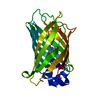
| ||||||||
|---|---|---|---|---|---|---|---|---|---|
| 1 |
| ||||||||
| Unit cell |
|
-Components
| #1: Protein | Mass: 25798.102 Da / Num. of mol.: 1 / Mutation: Q80R Source method: isolated from a genetically manipulated source Source: (gene. exp.) cfp marker plasmid pWM1009 (others) / Production host:  |
|---|---|
| #2: Water | ChemComp-HOH /  Mass: 18.015 Da / Num. of mol.: 166 / Source method: isolated from a natural source / Formula: H2O Mass: 18.015 Da / Num. of mol.: 166 / Source method: isolated from a natural source / Formula: H2O |
| Has protein modification | Y |
| Sequence details | RESIDUES 65THR, 66TRP AND 67GLY ARE MODIFIED TO MAKE THE CHROMOPHOR |
-Experimental details
-Experiment
| Experiment | Method: X-RAY DIFFRACTION / Number of used crystals: 1 |
|---|
- Sample preparation
| Crystal | Density Matthews: 2.15 Å3/Da / Density % sol: 42.92 % | ||||||||||||||||||||||||||||||||||||
|---|---|---|---|---|---|---|---|---|---|---|---|---|---|---|---|---|---|---|---|---|---|---|---|---|---|---|---|---|---|---|---|---|---|---|---|---|---|
| Crystal grow | Temperature: 298 K / Method: vapor diffusion, hanging drop / pH: 7 Details: PEG 1000, pH 7.0, VAPOR DIFFUSION, HANGING DROP, temperature 298K | ||||||||||||||||||||||||||||||||||||
| Crystal grow | *PLUS Temperature: 20 ℃ / Method: vapor diffusion, hanging drop | ||||||||||||||||||||||||||||||||||||
| Components of the solutions | *PLUS
|
-Data collection
| Diffraction | Mean temperature: 100 K |
|---|---|
| Diffraction source | Source: SYNCHROTRON / Site: MPG/DESY, HAMBURG  / Beamline: BW6 / Wavelength: 1.05 Å / Beamline: BW6 / Wavelength: 1.05 Å |
| Detector | Type: MARRESEARCH / Detector: CCD / Date: Mar 11, 2002 |
| Radiation | Monochromator: GRAPHITE / Protocol: SINGLE WAVELENGTH / Monochromatic (M) / Laue (L): M / Scattering type: x-ray |
| Radiation wavelength | Wavelength: 1.05 Å / Relative weight: 1 |
| Reflection | Resolution: 1.15→7.98 Å / Num. all: 79530 / Num. obs: 73514 / % possible obs: 92.4 % / Observed criterion σ(F): 2 / Observed criterion σ(I): 2 / Biso Wilson estimate: 11 Å2 |
| Reflection shell | Resolution: 1.15→1.22 Å / % possible all: 75.6 |
| Reflection | *PLUS Lowest resolution: 8 Å / Rmerge(I) obs: 0.039 |
| Reflection shell | *PLUS Lowest resolution: 1.17 Å / % possible obs: 84 % / Rmerge(I) obs: 0.247 |
- Processing
| Software |
| |||||||||||||||||||||||||
|---|---|---|---|---|---|---|---|---|---|---|---|---|---|---|---|---|---|---|---|---|---|---|---|---|---|---|
| Refinement | Method to determine structure: MOLECULAR REPLACEMENT / Resolution: 1.15→7.98 Å / Rfactor Rfree error: 0.003 / Isotropic thermal model: RESTRAINED / Cross valid method: THROUGHOUT / σ(F): 0 / Stereochemistry target values: Engh & Huber
| |||||||||||||||||||||||||
| Solvent computation | Solvent model: FLAT MODEL / Bsol: 68.4172 Å2 / ksol: 0.613 e/Å3 | |||||||||||||||||||||||||
| Displacement parameters | Biso mean: 15.5 Å2
| |||||||||||||||||||||||||
| Refine analyze |
| |||||||||||||||||||||||||
| Refinement step | Cycle: LAST / Resolution: 1.15→7.98 Å
| |||||||||||||||||||||||||
| Refine LS restraints |
| |||||||||||||||||||||||||
| LS refinement shell | Resolution: 1.15→1.22 Å / Rfactor Rfree error: 0.01 / Total num. of bins used: 6
| |||||||||||||||||||||||||
| Xplor file |
| |||||||||||||||||||||||||
| Refinement | *PLUS Lowest resolution: 8 Å / % reflection Rfree: 10 % / Rfactor Rfree: 0.228 / Rfactor Rwork: 0.215 | |||||||||||||||||||||||||
| Solvent computation | *PLUS | |||||||||||||||||||||||||
| Displacement parameters | *PLUS | |||||||||||||||||||||||||
| Refine LS restraints | *PLUS
|
