 Movie
Movie Controller


+ Open data

- Basic information
| Entry | Database: PDB / ID: 1ov3 | ||||||
|---|---|---|---|---|---|---|---|
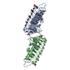
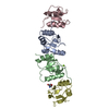
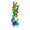
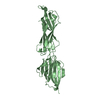
| Title | Structure of the p22phox-p47phox complex | ||||||
 Components Components |
| ||||||
 Keywords Keywords | Oxidoreductase activator / p47phox / p22phox / NADPH oxidase / complex | ||||||
| Function / homology |  Function and homology information Function and homology informationregulation of respiratory burst involved in inflammatory response / neutrophil-mediated killing of gram-positive bacterium / superoxide-generating NADPH oxidase activator activity / neutrophil-mediated killing of fungus / phagolysosome / cellular response to testosterone stimulus / Cross-presentation of particulate exogenous antigens (phagosomes) / superoxide-generating NAD(P)H oxidase activity / leukotriene metabolic process / NADPH oxidase complex ...regulation of respiratory burst involved in inflammatory response / neutrophil-mediated killing of gram-positive bacterium / superoxide-generating NADPH oxidase activator activity / neutrophil-mediated killing of fungus / phagolysosome / cellular response to testosterone stimulus / Cross-presentation of particulate exogenous antigens (phagosomes) / superoxide-generating NAD(P)H oxidase activity / leukotriene metabolic process / NADPH oxidase complex / response to yeast / respiratory burst involved in defense response / respiratory burst / phosphatidylinositol-3,4-bisphosphate binding / ROS and RNS production in phagocytes / hydrogen peroxide biosynthetic process / superoxide anion generation / protein targeting to membrane / superoxide metabolic process / Detoxification of Reactive Oxygen Species / RAC3 GTPase cycle / RAC2 GTPase cycle / RHO GTPases Activate NADPH Oxidases / cellular defense response / epithelial cell proliferation / RAC1 GTPase cycle / phosphatidylinositol binding / cellular response to glucose stimulus / SH3 domain binding / VEGFA-VEGFR2 Pathway / cytoplasmic side of plasma membrane / electron transfer activity / inflammatory response / innate immune response / neuronal cell body / dendrite / membrane / plasma membrane / cytosol / cytoplasm Similarity search - Function | ||||||
| Biological species |  Homo sapiens (human) Homo sapiens (human) | ||||||
| Method |  X-RAY DIFFRACTION / SYNCHROTRON / MOLECULAR REPLACEMENT / Resolution: 1.8 Å X-RAY DIFFRACTION / SYNCHROTRON / MOLECULAR REPLACEMENT / Resolution: 1.8 Å | ||||||
 Authors Authors | Groemping, Y. / Lapouge, K. / Smerdon, S.J. / Rittinger, K. | ||||||
 Citation Citation | Journal: Cell(Cambridge,Mass.) / Year: 2003 Title: Molecular basis of phosphorylation-induced activation of the NADPH oxidase Authors: Groemping, Y. / Lapouge, K. / Smerdon, S.J. / Rittinger, K. | ||||||
| History |
| ||||||
| Remark 300 | Biomolecule: The biologically active unit of p47phox is generated using amino acids 156-200 from ...Biomolecule: The biologically active unit of p47phox is generated using amino acids 156-200 from chain A and amino acids 201-285 from chain B. |
- Structure visualization
| Structure viewer | Molecule: MolmilJmol/JSmol |
|---|
- Downloads & links
-Download
| PDBx/mmCIF format | 1ov3.cif.gz | 72.8 KB | Display | PDBx/mmCIF format |
|---|---|---|---|---|
| PDB format | pdb1ov3.ent.gz | 54.5 KB | Display | PDB format |
| PDBx/mmJSON format | 1ov3.json.gz | Tree view | PDBx/mmJSON format | |
| Others |  Other downloads Other downloads |
-Validation report
| Arichive directory | https://data.pdbj.org/pub/pdb/validation_reports/ov/1ov3ftp://data.pdbj.org/pub/pdb/validation_reports/ov/1ov3 | HTTPS FTP |
|---|
-Related structure data
| Related structure data |  1ng2SC S: Starting model for refinement C: citing same article ( |
|---|---|
| Similar structure data |

-Links
 PDBj
PDBj


- Assembly
| Deposited unit | 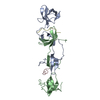
| ||||||||
|---|---|---|---|---|---|---|---|---|---|
| 1 |
| ||||||||
| 2 | 
| ||||||||
| Unit cell |
| ||||||||
| Components on special symmetry positions |
| ||||||||
| Details | The biologically active monomeric unit of p47phox is generated using amino acids 156-200 from chain A and amino acids 201-285 from chain B. |
-Components
| #1: Protein | Mass: 15396.137 Da / Num. of mol.: 2 / Fragment: Residues 156-285 Source method: isolated from a genetically manipulated source Source: (gene. exp.) Homo sapiens (human) / Gene: NCF1 / Plasmid: pGEX-6P1 / Species (production host): Escherichia coli / Production host:  #2: Protein/peptide | Mass: 2000.325 Da / Num. of mol.: 2 / Fragment: Residues 149-166 / Source method: obtained synthetically / Details: peptide synthesis / References: GenBank: 4557505 #3: Water | ChemComp-HOH / |  Mass: 18.015 Da / Num. of mol.: 207 / Source method: isolated from a natural source / Formula: H2O Mass: 18.015 Da / Num. of mol.: 207 / Source method: isolated from a natural source / Formula: H2O |
|---|
-Experimental details
-Experiment
| Experiment | Method: X-RAY DIFFRACTION / Number of used crystals: 1 |
|---|
- Sample preparation
| Crystal | Density Matthews: 2.49 Å3/Da / Density % sol: 50.54 % | |||||||||||||||||||||||||||||||||||||||||||||||||
|---|---|---|---|---|---|---|---|---|---|---|---|---|---|---|---|---|---|---|---|---|---|---|---|---|---|---|---|---|---|---|---|---|---|---|---|---|---|---|---|---|---|---|---|---|---|---|---|---|---|---|
| Crystal grow | Temperature: 291 K / Method: vapor diffusion, hanging drop / pH: 6.5 Details: 1 M Na-Citrate, 0.1 M Na-cacodylate, pH 6.5, VAPOR DIFFUSION, HANGING DROP, temperature 291K | |||||||||||||||||||||||||||||||||||||||||||||||||
| Crystal grow | *PLUS Temperature: 18 ℃ / pH: 7 / Method: vapor diffusion, hanging drop | |||||||||||||||||||||||||||||||||||||||||||||||||
| Components of the solutions | *PLUS
|
-Data collection
| Diffraction | Mean temperature: 100 K |
|---|---|
| Diffraction source | Source: SYNCHROTRON / Site: SRS  / Beamline: PX14.2 / Wavelength: 0.98 Å / Beamline: PX14.2 / Wavelength: 0.98 Å |
| Detector | Type: ADSC QUANTUM 4 / Detector: CCD / Date: Sep 3, 2002 / Details: Mirrors |
| Radiation | Monochromator: SI 111 / Protocol: SINGLE WAVELENGTH / Monochromatic (M) / Laue (L): M / Scattering type: x-ray |
| Radiation wavelength | Wavelength: 0.98 Å / Relative weight: 1 |
| Reflection | Resolution: 1.8→20 Å / Num. all: 226852 / Num. obs: 225037 / % possible obs: 99.2 % / Observed criterion σ(F): 0 / Observed criterion σ(I): 0 / Redundancy: 6.9 % / Rmerge(I) obs: 0.05 |
| Reflection shell | Resolution: 1.8→1.86 Å / Rmerge(I) obs: 0.37 / Mean I/σ(I) obs: 3.6 / Num. unique all: 3197 / % possible all: 99.1 |
| Reflection | *PLUS Lowest resolution: 20 Å / Rmerge(I) obs: 0.05 |
- Processing
| Software |
| |||||||||||||||||||||||||||||||||||||||||||||||||||||||||||||||||||||||||||||||||||||||||||||||||||||||||||||||||||||||||||||
|---|---|---|---|---|---|---|---|---|---|---|---|---|---|---|---|---|---|---|---|---|---|---|---|---|---|---|---|---|---|---|---|---|---|---|---|---|---|---|---|---|---|---|---|---|---|---|---|---|---|---|---|---|---|---|---|---|---|---|---|---|---|---|---|---|---|---|---|---|---|---|---|---|---|---|---|---|---|---|---|---|---|---|---|---|---|---|---|---|---|---|---|---|---|---|---|---|---|---|---|---|---|---|---|---|---|---|---|---|---|---|---|---|---|---|---|---|---|---|---|---|---|---|---|---|---|---|
| Refinement | Method to determine structure: MOLECULAR REPLACEMENT Starting model: 1NG2 Resolution: 1.8→20 Å / Cor.coef. Fo:Fc: 0.94 / Cor.coef. Fo:Fc free: 0.91 / SU B: 3.342 / SU ML: 0.106 / Cross valid method: THROUGHOUT / σ(F): 0 / ESU R: 0.143 / ESU R Free: 0.142 / Stereochemistry target values: MAXIMUM LIKELIHOOD
| |||||||||||||||||||||||||||||||||||||||||||||||||||||||||||||||||||||||||||||||||||||||||||||||||||||||||||||||||||||||||||||
| Solvent computation | Ion probe radii: 0.8 Å / Shrinkage radii: 0.8 Å / VDW probe radii: 1.4 Å / Solvent model: BABINET MODEL WITH MASK | |||||||||||||||||||||||||||||||||||||||||||||||||||||||||||||||||||||||||||||||||||||||||||||||||||||||||||||||||||||||||||||
| Displacement parameters | Biso mean: 21.218 Å2
| |||||||||||||||||||||||||||||||||||||||||||||||||||||||||||||||||||||||||||||||||||||||||||||||||||||||||||||||||||||||||||||
| Refinement step | Cycle: LAST / Resolution: 1.8→20 Å
| |||||||||||||||||||||||||||||||||||||||||||||||||||||||||||||||||||||||||||||||||||||||||||||||||||||||||||||||||||||||||||||
| Refine LS restraints |
| |||||||||||||||||||||||||||||||||||||||||||||||||||||||||||||||||||||||||||||||||||||||||||||||||||||||||||||||||||||||||||||
| LS refinement shell | Resolution: 1.802→1.848 Å / Total num. of bins used: 20
| |||||||||||||||||||||||||||||||||||||||||||||||||||||||||||||||||||||||||||||||||||||||||||||||||||||||||||||||||||||||||||||
| Refinement TLS params. | Method: refined / Refine-ID: X-RAY DIFFRACTION
| |||||||||||||||||||||||||||||||||||||||||||||||||||||||||||||||||||||||||||||||||||||||||||||||||||||||||||||||||||||||||||||
| Refinement TLS group |
| |||||||||||||||||||||||||||||||||||||||||||||||||||||||||||||||||||||||||||||||||||||||||||||||||||||||||||||||||||||||||||||
| Software | *PLUS Version: 5 / Classification: refinement | |||||||||||||||||||||||||||||||||||||||||||||||||||||||||||||||||||||||||||||||||||||||||||||||||||||||||||||||||||||||||||||
| Refinement | *PLUS Highest resolution: 1.8 Å / Lowest resolution: 20 Å / % reflection Rfree: 5 % / Rfactor Rwork: 0.232 | |||||||||||||||||||||||||||||||||||||||||||||||||||||||||||||||||||||||||||||||||||||||||||||||||||||||||||||||||||||||||||||
| Solvent computation | *PLUS | |||||||||||||||||||||||||||||||||||||||||||||||||||||||||||||||||||||||||||||||||||||||||||||||||||||||||||||||||||||||||||||
| Displacement parameters | *PLUS | |||||||||||||||||||||||||||||||||||||||||||||||||||||||||||||||||||||||||||||||||||||||||||||||||||||||||||||||||||||||||||||
| Refine LS restraints | *PLUS
|
