 Movie
Movie Controller


+ Open data

- Basic information
| Entry | Database: PDB / ID: 1oul | ||||||
|---|---|---|---|---|---|---|---|
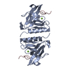
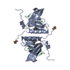
| Title | Structure of the AAA+ protease delivery protein SspB | ||||||
 Components Components | Stringent starvation protein B homolog | ||||||
 Keywords Keywords | TRANSPORT PROTEIN / ssrA peptide binding protein / homodimer | ||||||
| Function / homology |  Function and homology information Function and homology information | ||||||
| Biological species |  Haemophilus influenzae (bacteria) Haemophilus influenzae (bacteria) | ||||||
| Method |  X-RAY DIFFRACTION / SYNCHROTRON / MAD / Resolution: 2.2 Å X-RAY DIFFRACTION / SYNCHROTRON / MAD / Resolution: 2.2 Å | ||||||
 Authors Authors | Levchenko, I. / Grant, R.A. / Wah, D.A. / Sauer, R.T. / Baker, T.A. | ||||||
 Citation Citation | Journal: Mol.Cell / Year: 2003 Title: Structure of a delivery protein for an AAA+ protease in complex with a peptide degradation tag Authors: Levchenko, I. / Grant, R.A. / Wah, D.A. / Sauer, R.T. / Baker, T.A. #1: Journal: Science / Year: 2000Title: A specificity-enhancing factor for the ClpXP degradation machine Authors: Levchenko, I. / Siedel, M. / Sauer, R.T. / Baker, T.A. | ||||||
| History |
|
- Structure visualization
| Structure viewer | Molecule: MolmilJmol/JSmol |
|---|
- Downloads & links
-Download
| PDBx/mmCIF format | 1oul.cif.gz | 61.3 KB | Display | PDBx/mmCIF format |
|---|---|---|---|---|
| PDB format | pdb1oul.ent.gz | 45.9 KB | Display | PDB format |
| PDBx/mmJSON format | 1oul.json.gz | Tree view | PDBx/mmJSON format | |
| Others |  Other downloads Other downloads |
-Validation report
| Arichive directory | https://data.pdbj.org/pub/pdb/validation_reports/ou/1oulftp://data.pdbj.org/pub/pdb/validation_reports/ou/1oul | HTTPS FTP |
|---|
-Related structure data


-Links
 PDBj
PDBj- Assembly
| Deposited unit | 
| ||||||||
|---|---|---|---|---|---|---|---|---|---|
| 1 |
| ||||||||
| Unit cell |
| ||||||||
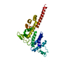
| Details | the biological homodimers consists of chains A and B together |
-Components
| #1: Protein | Mass: 14801.002 Da / Num. of mol.: 2 / Fragment: residues 1-129 Source method: isolated from a genetically manipulated source Source: (gene. exp.) Haemophilus influenzae (bacteria) / Gene: SSPB / Production host: #2: Water | ChemComp-HOH / |  Mass: 18.015 Da / Num. of mol.: 65 / Source method: isolated from a natural source / Formula: H2O Mass: 18.015 Da / Num. of mol.: 65 / Source method: isolated from a natural source / Formula: H2OHas protein modification | Y | |
|---|
-Experimental details
-Experiment
| Experiment | Method: X-RAY DIFFRACTION / Number of used crystals: 1 |
|---|
- Sample preparation
| Crystal | Density Matthews: 3.35 Å3/Da / Density % sol: 63 % | ||||||||||||||||||||||||||||||
|---|---|---|---|---|---|---|---|---|---|---|---|---|---|---|---|---|---|---|---|---|---|---|---|---|---|---|---|---|---|---|---|
| Crystal grow | Temperature: 298 K / Method: vapor diffusion, sitting drop / pH: 6.3 Details: sodium formate, sodium cacodylate, ammonium sulfate, pH 6.3, VAPOR DIFFUSION, SITTING DROP, temperature 298K | ||||||||||||||||||||||||||||||
| Crystal grow | *PLUS Temperature: 20 ℃ / pH: 5.6 / Method: vapor diffusion, sitting drop | ||||||||||||||||||||||||||||||
| Components of the solutions | *PLUS
|
-Data collection
| Diffraction | Mean temperature: 113 K | ||||||||||||
|---|---|---|---|---|---|---|---|---|---|---|---|---|---|
| Diffraction source | Source: SYNCHROTRON / Site: NSLS  / Beamline: X4A / Wavelength: 0.97898, 0.97939, 0.96866 / Beamline: X4A / Wavelength: 0.97898, 0.97939, 0.96866 | ||||||||||||
| Detector | Type: ADSC QUANTUM 4 / Detector: CCD / Date: Jun 26, 2002 | ||||||||||||
| Radiation | Monochromator: two crystal monochromator / Protocol: MAD / Monochromatic (M) / Laue (L): M / Scattering type: x-ray | ||||||||||||
| Radiation wavelength |
| ||||||||||||
| Reflection | Resolution: 2.1→20 Å / Num. all: 23481 / Num. obs: 22666 / % possible obs: 99.9 % / Observed criterion σ(F): 0 / Observed criterion σ(I): 0 / Biso Wilson estimate: 36.4 Å2 / Rsym value: 0.092 | ||||||||||||
| Reflection shell | Resolution: 2.1→2.17 Å / % possible all: 91.8 |
- Processing
| Software |
| |||||||||||||||||||||||||
|---|---|---|---|---|---|---|---|---|---|---|---|---|---|---|---|---|---|---|---|---|---|---|---|---|---|---|
| Refinement | Method to determine structure: MAD / Resolution: 2.2→19.72 Å / Rfactor Rfree error: 0.005 / Data cutoff high rms absF: 236909.41 / Isotropic thermal model: RESTRAINED / Cross valid method: THROUGHOUT / σ(F): 2 / Stereochemistry target values: Engh & Huber Details: Reflection count for refinement statistics are approximately twice as large as the scaling reflection count because the refinement statistics do not reflect the merging of Bjvoet pairs
| |||||||||||||||||||||||||
| Solvent computation | Solvent model: FLAT MODEL / Bsol: 43.283 Å2 / ksol: 0.362347 e/Å3 | |||||||||||||||||||||||||
| Displacement parameters | Biso mean: 49.5 Å2
| |||||||||||||||||||||||||
| Refine analyze |
| |||||||||||||||||||||||||
| Refinement step | Cycle: LAST / Resolution: 2.2→19.72 Å
| |||||||||||||||||||||||||
| Refine LS restraints |
| |||||||||||||||||||||||||
| LS refinement shell | Resolution: 2.2→2.34 Å / Rfactor Rfree error: 0.016 / Total num. of bins used: 6
| |||||||||||||||||||||||||
| Xplor file |
| |||||||||||||||||||||||||
| Refine LS restraints | *PLUS
|
