 Movie
Movie Controller


+ Open data

- Basic information
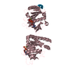
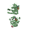
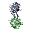
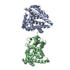
| Entry | Database: PDB / ID: 1otj | ||||||
|---|---|---|---|---|---|---|---|
| Title | Crystal structure of APO (iron-free) TauD | ||||||
 Components Components | Alpha-Ketoglutarate-Dependent Taurine Dioxygenase | ||||||
 Keywords Keywords | OXIDOREDUCTASE / Jelly roll motif / alpha ketoglutarate-dependent dioxygenase / taurine | ||||||
| Function / homology |  Function and homology information Function and homology informationtaurine catabolic process / taurine dioxygenase complex / taurine dioxygenase / taurine dioxygenase activity / sulfur compound metabolic process / L-ascorbic acid binding / ferrous iron binding / protein homotetramerization / identical protein binding / cytoplasm Similarity search - Function | ||||||
| Biological species |  | ||||||
| Method |  X-RAY DIFFRACTION / Direct methods, MAD / Resolution: 1.9 Å X-RAY DIFFRACTION / Direct methods, MAD / Resolution: 1.9 Å | ||||||
 Authors Authors | O'Brien, J.R. / Schuller, D.J. / Yang, V.S. / Dillard, B.D. / Lanzilotta, W.N. | ||||||
 Citation Citation | Journal: Biochemistry / Year: 2003 Title: Substrate-induced conformational changes in Escherichia coli taurine/alpha-ketoglutarate dioxygenase and insight into the oligomeric structure Authors: O'Brien, J.R. / Schuller, D.J. / Yang, V.S. / Dillard, B.D. / Lanzilotta, W.N. | ||||||
| History |
|
- Structure visualization
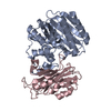
| Structure viewer | Molecule: MolmilJmol/JSmol |
|---|
- Downloads & links
-Download
| PDBx/mmCIF format | 1otj.cif.gz | 247.3 KB | Display | PDBx/mmCIF format |
|---|---|---|---|---|
| PDB format | pdb1otj.ent.gz | 200.9 KB | Display | PDB format |
| PDBx/mmJSON format | 1otj.json.gz | Tree view | PDBx/mmJSON format | |
| Others |  Other downloads Other downloads |
-Validation report
| Arichive directory | https://data.pdbj.org/pub/pdb/validation_reports/ot/1otjftp://data.pdbj.org/pub/pdb/validation_reports/ot/1otj | HTTPS FTP |
|---|
-Related structure data


-Links
 PDBj
PDBj- Assembly
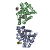
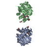
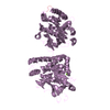
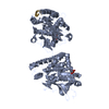
| Deposited unit | 
| ||||||||
|---|---|---|---|---|---|---|---|---|---|
| 1 | 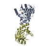
| ||||||||
| 2 | 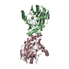
| ||||||||
| Unit cell |
| ||||||||
| Details | Two biological dimers are found in the crystallographic tetramer. They are monomers A & E, and B & D. |
-Components
| #1: Protein | Mass: 32453.467 Da / Num. of mol.: 4 Source method: isolated from a genetically manipulated source Source: (gene. exp.) #2: Chemical |   Mass: 35.453 Da / Num. of mol.: 3 / Source method: obtained synthetically / Formula: Cl Mass: 35.453 Da / Num. of mol.: 3 / Source method: obtained synthetically / Formula: Cl#3: Chemical | 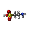  Mass: 125.147 Da / Num. of mol.: 3 / Source method: obtained synthetically / Formula: C2H7NO3S Mass: 125.147 Da / Num. of mol.: 3 / Source method: obtained synthetically / Formula: C2H7NO3S#4: Water | ChemComp-HOH / |  Mass: 18.015 Da / Num. of mol.: 710 / Source method: isolated from a natural source / Formula: H2O Mass: 18.015 Da / Num. of mol.: 710 / Source method: isolated from a natural source / Formula: H2O |
|---|
-Experimental details
-Experiment
| Experiment | Method: X-RAY DIFFRACTION / Number of used crystals: 1 |
|---|
- Sample preparation
| Crystal | Density Matthews: 2.4 Å3/Da / Density % sol: 48.4 % | ||||||||||||||||||||||||||||||
|---|---|---|---|---|---|---|---|---|---|---|---|---|---|---|---|---|---|---|---|---|---|---|---|---|---|---|---|---|---|---|---|
| Crystal grow | Temperature: 295 K / Method: batch / pH: 8 Details: PEG 4000, Isopropanol, Sodium citrate, pH 8.0, Batch, temperature 295K | ||||||||||||||||||||||||||||||
| Crystal grow | *PLUS Temperature: 22 ℃ / Method: batch method | ||||||||||||||||||||||||||||||
| Components of the solutions | *PLUS
|
-Data collection
| Diffraction | Mean temperature: 93 K |
|---|---|
| Diffraction source | Source: ROTATING ANODE / Type: RIGAKU RU200 / Wavelength: 1.54 Å |
| Detector | Type: RIGAKU RAXIS IIC / Detector: IMAGE PLATE / Date: Sep 21, 2002 / Details: Osmic Blue confocal mirrors |
| Radiation | Monochromator: Confocal Mirrors / Protocol: SINGLE WAVELENGTH / Monochromatic (M) / Laue (L): M / Scattering type: x-ray |
| Radiation wavelength | Wavelength: 1.54 Å / Relative weight: 1 |
| Reflection | Resolution: 1.9→50 Å / Num. all: 105897 / Num. obs: 99459 / % possible obs: 94 % / Observed criterion σ(F): 2 / Observed criterion σ(I): 2 / Redundancy: 5.5 % / Biso Wilson estimate: 14.2 Å2 / Rmerge(I) obs: 0.094 / Rsym value: 0.049 / Net I/σ(I): 15.4 |
| Reflection shell | Resolution: 1.9→1.97 Å / Redundancy: 3.2 % / Rmerge(I) obs: 0.27 / Mean I/σ(I) obs: 3.1 / Num. unique all: 600 / Rsym value: 0.34 / % possible all: 93.5 |
| Reflection | *PLUS Highest resolution: 1.9 Å / % possible obs: 96.9 % / Rmerge(I) obs: 0.049 |
| Reflection shell | *PLUS Highest resolution: 1.9 Å / % possible obs: 78.9 % / Rmerge(I) obs: 0.334 |
- Processing
| Software |
| ||||||||||||||||||||||||||||||||||||||||||||||||||||||||||||||||||||||||||||||||
|---|---|---|---|---|---|---|---|---|---|---|---|---|---|---|---|---|---|---|---|---|---|---|---|---|---|---|---|---|---|---|---|---|---|---|---|---|---|---|---|---|---|---|---|---|---|---|---|---|---|---|---|---|---|---|---|---|---|---|---|---|---|---|---|---|---|---|---|---|---|---|---|---|---|---|---|---|---|---|---|---|---|
| Refinement | Method to determine structure: Direct methods, MAD / Resolution: 1.9→50 Å / Isotropic thermal model: Anisotropic / Cross valid method: THROUGHOUT / σ(F): 2 / σ(I): 2 / Stereochemistry target values: Engh & Huber
| ||||||||||||||||||||||||||||||||||||||||||||||||||||||||||||||||||||||||||||||||
| Displacement parameters | Biso mean: 34.3 Å2
| ||||||||||||||||||||||||||||||||||||||||||||||||||||||||||||||||||||||||||||||||
| Refinement step | Cycle: LAST / Resolution: 1.9→50 Å
| ||||||||||||||||||||||||||||||||||||||||||||||||||||||||||||||||||||||||||||||||
| Refine LS restraints |
| ||||||||||||||||||||||||||||||||||||||||||||||||||||||||||||||||||||||||||||||||
| Xplor file |
| ||||||||||||||||||||||||||||||||||||||||||||||||||||||||||||||||||||||||||||||||
| Refinement | *PLUS Num. reflection Rfree: 5034 / Rfactor Rfree: 0.254 | ||||||||||||||||||||||||||||||||||||||||||||||||||||||||||||||||||||||||||||||||
| Solvent computation | *PLUS | ||||||||||||||||||||||||||||||||||||||||||||||||||||||||||||||||||||||||||||||||
| Displacement parameters | *PLUS | ||||||||||||||||||||||||||||||||||||||||||||||||||||||||||||||||||||||||||||||||
| Refine LS restraints | *PLUS
|
