 Movie
Movie Controller

[English] 日本語
 Yorodumi
- PDB-1omz: crystal structure of mouse alpha-1,4-N-acetylhexosaminyltransfera... -
+ Open data

- Basic information
| Entry | Database: PDB / ID: 1omz | ||||||
|---|---|---|---|---|---|---|---|
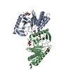
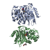
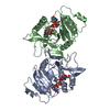
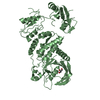
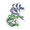
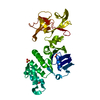
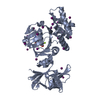
| Title | crystal structure of mouse alpha-1,4-N-acetylhexosaminyltransferase (EXTL2) in complex with UDPGalNAc | ||||||
 Components Components | Alpha-1,4-N-acetylhexosaminyltransferase EXTL2 | ||||||
 Keywords Keywords | TRANSFERASE / Rossmann fold / DXD motif | ||||||
| Function / homology |  Function and homology information Function and homology informationalpha-1,4-N-acetylgalactosaminyltransferase activity / glucuronylgalactosylproteoglycan 4-beta-N-acetylgalactosaminyltransferase activity / UDP-N-acetylgalactosamine metabolic process / glucuronosyl-galactosyl-proteoglycan 4-alpha-N-acetylglucosaminyltransferase / glucuronyl-galactosyl-proteoglycan 4-alpha-N-acetylglucosaminyltransferase activity / HS-GAG biosynthesis / N-acetylglucosamine metabolic process / glycosaminoglycan binding / manganese ion binding / endoplasmic reticulum membrane ...alpha-1,4-N-acetylgalactosaminyltransferase activity / glucuronylgalactosylproteoglycan 4-beta-N-acetylgalactosaminyltransferase activity / UDP-N-acetylgalactosamine metabolic process / glucuronosyl-galactosyl-proteoglycan 4-alpha-N-acetylglucosaminyltransferase / glucuronyl-galactosyl-proteoglycan 4-alpha-N-acetylglucosaminyltransferase activity / HS-GAG biosynthesis / N-acetylglucosamine metabolic process / glycosaminoglycan binding / manganese ion binding / endoplasmic reticulum membrane / endoplasmic reticulum / nucleoplasm / cytosol Similarity search - Function | ||||||
| Biological species |  | ||||||
| Method |  X-RAY DIFFRACTION / MOLECULAR REPLACEMENT / Resolution: 2.1 Å X-RAY DIFFRACTION / MOLECULAR REPLACEMENT / Resolution: 2.1 Å | ||||||
 Authors Authors | Pedersen, L.C. / Dong, J. / Taniguchi, F. / Kitagawa, H. / Krahn, J.M. / Pedersen, L.G. / Sugahara, K. / Negishi, M. | ||||||
 Citation Citation | Journal: J.Biol.Chem. / Year: 2003 Title: Crystal structure of an alpha-1,4-N-acetylhexosaminyltransferase (EXTL2), a member of the exostosin gene family involved in heparan sulfate biosynthesis Authors: Pedersen, L.C. / Dong, J. / Taniguchi, F. / Kitagawa, H. / Krahn, J.M. / Pedersen, L.G. / Sugahara, K. / Negishi, M. | ||||||
| History |
| ||||||
| Remark 300 | BIOMOLECULE THIS ENTRY CONTAINS THE CRYSTALLOGRAPHIC ASYMMETRIC UNIT WHICH CONSISTS OF 2 CHAIN(S). ...BIOMOLECULE THIS ENTRY CONTAINS THE CRYSTALLOGRAPHIC ASYMMETRIC UNIT WHICH CONSISTS OF 2 CHAIN(S). THE BIOLOGICAL UNIT IS NOT KNOWN. | ||||||
| Remark 999 | SEQUENCE Although the fusion protein incodes residues 38-330, the exact N-termini is not known due ...SEQUENCE Although the fusion protein incodes residues 38-330, the exact N-termini is not known due to use of a non-specific protease to cleave of the maltose binding protein. However residues 62-327 are visible in the electron density. |
- Structure visualization
| Structure viewer | Molecule: MolmilJmol/JSmol |
|---|
- Downloads & links
-Download
| PDBx/mmCIF format | 1omz.cif.gz | 123.7 KB | Display | PDBx/mmCIF format |
|---|---|---|---|---|
| PDB format | pdb1omz.ent.gz | 94.1 KB | Display | PDB format |
| PDBx/mmJSON format | 1omz.json.gz | Tree view | PDBx/mmJSON format | |
| Others |  Other downloads Other downloads |
-Validation report
| Arichive directory | https://data.pdbj.org/pub/pdb/validation_reports/om/1omzftp://data.pdbj.org/pub/pdb/validation_reports/om/1omz | HTTPS FTP |
|---|
-Related structure data
| Related structure data | 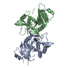 1omxSC 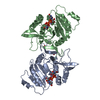 1on6C  1on8C S: Starting model for refinement C: citing same article ( |
|---|---|
| Similar structure data |


-Links
 PDBj
PDBj

- Assembly
| Deposited unit | 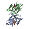
| ||||||||
|---|---|---|---|---|---|---|---|---|---|
| 1 |
| ||||||||
| 2 | 
| ||||||||
| Unit cell |
|
-Components
| #1: Protein | Mass: 33396.312 Da / Num. of mol.: 2 / Fragment: catalytic domain Source method: isolated from a genetically manipulated source Source: (gene. exp.)  References: UniProt: Q9ES89, Transferases; Glycosyltransferases; Hexosyltransferases #2: Chemical |   Mass: 54.938 Da / Num. of mol.: 2 / Source method: obtained synthetically / Formula: Mn Mass: 54.938 Da / Num. of mol.: 2 / Source method: obtained synthetically / Formula: Mn#3: Chemical |   Mass: 607.354 Da / Num. of mol.: 2 / Source method: obtained synthetically / Formula: C17H27N3O17P2 Mass: 607.354 Da / Num. of mol.: 2 / Source method: obtained synthetically / Formula: C17H27N3O17P2#4: Chemical |   Mass: 62.068 Da / Num. of mol.: 2 / Source method: obtained synthetically / Formula: C2H6O2 Mass: 62.068 Da / Num. of mol.: 2 / Source method: obtained synthetically / Formula: C2H6O2#5: Water | ChemComp-HOH / |  Mass: 18.015 Da / Num. of mol.: 249 / Source method: isolated from a natural source / Formula: H2O Mass: 18.015 Da / Num. of mol.: 249 / Source method: isolated from a natural source / Formula: H2OHas protein modification | Y | |
|---|
-Experimental details
-Experiment
| Experiment | Method: X-RAY DIFFRACTION / Number of used crystals: 1 |
|---|
- Sample preparation
| Crystal | Density Matthews: 2.51 Å3/Da / Density % sol: 50.91 % | ||||||||||||||||||||||||||||||||||||||||||
|---|---|---|---|---|---|---|---|---|---|---|---|---|---|---|---|---|---|---|---|---|---|---|---|---|---|---|---|---|---|---|---|---|---|---|---|---|---|---|---|---|---|---|---|
| Crystal grow | Temperature: 277 K / Method: vapor diffusion, hanging drop / pH: 7.5 Details: PEG3000, magnesium chloride, cacodylate, pH 7.5, VAPOR DIFFUSION, HANGING DROP, temperature 277K | ||||||||||||||||||||||||||||||||||||||||||
| Crystal grow | *PLUS | ||||||||||||||||||||||||||||||||||||||||||
| Components of the solutions | *PLUS
|
-Data collection
| Diffraction | Mean temperature: 100 K |
|---|---|
| Diffraction source | Source: ROTATING ANODE / Type: RIGAKU RU300 / Wavelength: 1.5418 Å |
| Detector | Type: RIGAKU RAXIS IV / Detector: IMAGE PLATE / Date: Apr 15, 2002 / Details: mirrors |
| Radiation | Monochromator: mirrors / Protocol: SINGLE WAVELENGTH / Monochromatic (M) / Laue (L): M / Scattering type: x-ray |
| Radiation wavelength | Wavelength: 1.5418 Å / Relative weight: 1 |
| Reflection | Resolution: 2.1→50 Å / Num. obs: 39701 / % possible obs: 98.9 % / Observed criterion σ(F): -3 / Observed criterion σ(I): -3 / Redundancy: 5 % / Biso Wilson estimate: 13.5 Å2 / Rsym value: 0.097 / Net I/σ(I): 17.7 |
| Reflection shell | Resolution: 2.1→2.17 Å / Redundancy: 3.2 % / Mean I/σ(I) obs: 2.8 / Num. unique all: 3720 / Rsym value: 0.396 / % possible all: 95 |
| Reflection | *PLUS Lowest resolution: 50 Å / Num. measured all: 197735 / Rmerge(I) obs: 0.097 |
| Reflection shell | *PLUS % possible obs: 95 % / Rmerge(I) obs: 0.396 |
- Processing
| Software |
| ||||||||||||||||||||||||||||||||||||
|---|---|---|---|---|---|---|---|---|---|---|---|---|---|---|---|---|---|---|---|---|---|---|---|---|---|---|---|---|---|---|---|---|---|---|---|---|---|
| Refinement | Method to determine structure: MOLECULAR REPLACEMENT Starting model: 1OMX Resolution: 2.1→19.88 Å / Rfactor Rfree error: 0.005 / Isotropic thermal model: RESTRAINED / Cross valid method: THROUGHOUT / σ(F): 0 / Stereochemistry target values: Engh & Huber
| ||||||||||||||||||||||||||||||||||||
| Solvent computation | Solvent model: FLAT MODEL / Bsol: 53.1872 Å2 / ksol: 0.358562 e/Å3 | ||||||||||||||||||||||||||||||||||||
| Displacement parameters | Biso mean: 29 Å2
| ||||||||||||||||||||||||||||||||||||
| Refine analyze | Luzzati coordinate error free: 0.29 Å / Luzzati sigma a free: 0.29 Å | ||||||||||||||||||||||||||||||||||||
| Refinement step | Cycle: LAST / Resolution: 2.1→19.88 Å
| ||||||||||||||||||||||||||||||||||||
| Refine LS restraints |
| ||||||||||||||||||||||||||||||||||||
| LS refinement shell | Resolution: 2.1→2.23 Å / Rfactor Rfree error: 0.018 / Total num. of bins used: 6
| ||||||||||||||||||||||||||||||||||||
| Xplor file |
| ||||||||||||||||||||||||||||||||||||
| Software | *PLUS Classification: refinement | ||||||||||||||||||||||||||||||||||||
| Refinement | *PLUS % reflection Rfree: 5 % / Highest resolution: 2.1 Å / Lowest resolution: 50 Å | ||||||||||||||||||||||||||||||||||||
| Solvent computation | *PLUS | ||||||||||||||||||||||||||||||||||||
| Displacement parameters | *PLUS | ||||||||||||||||||||||||||||||||||||
| Refine LS restraints | *PLUS
|
