- PDB-1oiz: The Molecular Basis of Vitamin E Retention: Structure of Human Al... -
+
Open data
ID or keywords:
Loading...
-
Basic information
Entry
Database: PDB / ID: 1oiz
Title
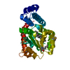
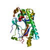
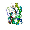
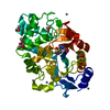
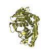
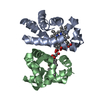
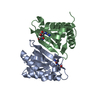
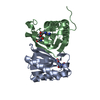
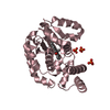
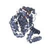
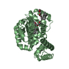
The Molecular Basis of Vitamin E Retention: Structure of Human Alpha-Tocopherol Transfer Protein
Components
ALPHA-TOCOPHEROL TRANSFER PROTEIN
Keywords
TRANSPORT / ATAXIA / AVED / TRANSFER PROTEIN / TOCOPHEROL / VITAMIN E
Function / homology
Function and homology information
Vitamin E transport / vitamin E binding / vitamin E metabolic process / vitamin transport / lipid transfer activity / intermembrane lipid transfer / negative regulation of establishment of blood-brain barrier / positive regulation of amyloid-beta clearance / phosphatidylinositol-3,4-bisphosphate binding / phosphatidylinositol bisphosphate binding ...Vitamin E transport / vitamin E binding / vitamin E metabolic process / vitamin transport / lipid transfer activity / intermembrane lipid transfer / negative regulation of establishment of blood-brain barrier / positive regulation of amyloid-beta clearance / phosphatidylinositol-3,4-bisphosphate binding / phosphatidylinositol bisphosphate binding / embryonic placenta development / phosphatidylinositol-4,5-bisphosphate binding / lipid metabolic process / response to toxic substance / late endosome / cytosol Similarity search - Function
N-terminal domain of phosphatidylinositol transfer protein sec14p / Phosphatidylinositol Transfer Protein Sec14p / CRAL-TRIO lipid binding domain / CRAL/TRIO, N-terminal domain / CRAL/TRIO, N-terminal domain / CRAL/TRIO, N-terminal domain / CRAL/TRIO, N-terminal domain superfamily / CRAL/TRIO domain / CRAL-TRIO lipid binding domain profile. / Domain in homologues of a S. cerevisiae phosphatidylinositol transfer protein (Sec14p) ...N-terminal domain of phosphatidylinositol transfer protein sec14p / Phosphatidylinositol Transfer Protein Sec14p / CRAL-TRIO lipid binding domain / CRAL/TRIO, N-terminal domain / CRAL/TRIO, N-terminal domain / CRAL/TRIO, N-terminal domain / CRAL/TRIO, N-terminal domain superfamily / CRAL/TRIO domain / CRAL-TRIO lipid binding domain profile. / Domain in homologues of a S. cerevisiae phosphatidylinositol transfer protein (Sec14p) / CRAL-TRIO lipid binding domain / CRAL-TRIO lipid binding domain superfamily / Helicase, Ruva Protein; domain 3 / Orthogonal Bundle / 3-Layer(aba) Sandwich / Mainly Alpha / Alpha Beta Similarity search - Domain/homology
Mass: 18.015 Da / Num. of mol.: 294 / Source method: isolated from a natural source / Formula: H2O
Compound details
FUNCTION: BINDS ALPHA-TOCOPHEROL AND ENHANCES ITS TRANSFER BETWEEN SEPARATE MEMBRANES. DISEASE: ...FUNCTION: BINDS ALPHA-TOCOPHEROL AND ENHANCES ITS TRANSFER BETWEEN SEPARATE MEMBRANES. DISEASE: DEFECTS IN TTPA ARE THE CAUSE OF ATAXIA WITH ISOLATED VITAMIN E DEFICIENCY
Has protein modification
N
-
Experimental details
-
Experiment
Experiment
Method: X-RAY DIFFRACTION / Number of used crystals: 1
-
Sample preparation
Crystal
Density Matthews: 2.69 Å3/Da / Density % sol: 54.28 %
 Movie
Movie Controller

 Yorodumi
 Open data
 Basic information
 Components
 Keywords
 Function and homology information
 HOMO SAPIENS (human)
 X-RAY DIFFRACTION /
  Authors
 Citation
 Structure visualization
 Downloads & links
 Other downloads
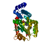


 PDBj
PDBj Assembly
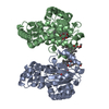


 Mass: 352.508 Da / Num. of mol.: 6 / Source method: obtained synthetically / Formula: C21H36O4 / Comment: detergent*YM
 Mass: 18.015 Da / Num. of mol.: 294 / Source method: isolated from a natural source / Formula: H2O
 Sample preparation
 / Beamline: X06SA / Wavelength: 0.9791
  Processing