 Movie
Movie Controller


+ Open data

- Basic information
| Entry | Database: PDB / ID: 1oej | ||||||
|---|---|---|---|---|---|---|---|
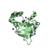
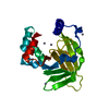
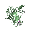
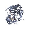
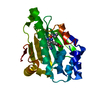
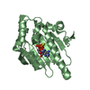
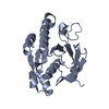
| Title | YodA from Escherichia coli crystallised with no added ions | ||||||
 Components Components | HYPOTHETICAL PROTEIN YODA | ||||||
 Keywords Keywords | STRESS PROTEIN/LIPOCALIN / STRESS PROTEIN / LIPOCALIN / YODA / STRESS PROTEIN-LIPOCALIN complex | ||||||
| Function / homology |  Function and homology information Function and homology informationcellular response to zinc ion starvation / intracellular zinc ion homeostasis / cadmium ion binding / cellular response to cadmium ion / cellular response to hydrogen peroxide / outer membrane-bounded periplasmic space / zinc ion binding / metal ion binding / cytosol Similarity search - Function | ||||||
| Biological species |  | ||||||
| Method |  X-RAY DIFFRACTION / SYNCHROTRON / MOLECULAR REPLACEMENT / Resolution: 1.81 Å X-RAY DIFFRACTION / SYNCHROTRON / MOLECULAR REPLACEMENT / Resolution: 1.81 Å | ||||||
 Authors Authors | David, G. / Blondeau, K. / Renouard, M. / Penel, S. / Lewit-Bentley, A. | ||||||
 Citation Citation | Journal: J.Biol.Chem. / Year: 2003 Title: Yoda from Escherichia Coli is a Metal-Binding, Lipocalin-Like Protein Authors: David, G. / Blondeau, K. / Schiltz, M. / Penel, S. / Lewit-Bentley, A. | ||||||
| History |
|
- Structure visualization
| Structure viewer | Molecule: MolmilJmol/JSmol |
|---|
- Downloads & links
-Download
| PDBx/mmCIF format | 1oej.cif.gz | 52.4 KB | Display | PDBx/mmCIF format |
|---|---|---|---|---|
| PDB format | pdb1oej.ent.gz | 37.2 KB | Display | PDB format |
| PDBx/mmJSON format | 1oej.json.gz | Tree view | PDBx/mmJSON format | |
| Others |  Other downloads Other downloads |
-Validation report
| Arichive directory | https://data.pdbj.org/pub/pdb/validation_reports/oe/1oejftp://data.pdbj.org/pub/pdb/validation_reports/oe/1oej | HTTPS FTP |
|---|
-Related structure data
| Related structure data | 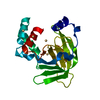 1oeeSC  1oekC S: Starting model for refinement C: citing same article ( |
|---|---|
| Similar structure data |


-Links
 PDBj
PDBj

- Assembly
| Deposited unit | 
| ||||||||
|---|---|---|---|---|---|---|---|---|---|
| 1 |
| ||||||||
| Unit cell |
|
-Components
| #1: Protein | Mass: 22373.879 Da / Num. of mol.: 1 / Source method: isolated from a natural source / Source: (natural) |
|---|---|
| #2: Chemical | ChemComp-NI /   Mass: 58.693 Da / Num. of mol.: 1 / Source method: obtained synthetically / Formula: Ni Mass: 58.693 Da / Num. of mol.: 1 / Source method: obtained synthetically / Formula: Ni |
| #3: Water | ChemComp-HOH /  Mass: 18.015 Da / Num. of mol.: 101 / Source method: isolated from a natural source / Formula: H2O Mass: 18.015 Da / Num. of mol.: 101 / Source method: isolated from a natural source / Formula: H2O |
| Has protein modification | Y |
| Sequence details | THE SEQUENCE: MAIRLYKLAVALGVFIVSAPAFS THAT IS INCLUDED THE SWISPROT RECORD UPSTREAM OF THE SEQUENCE ...THE SEQUENCE: MAIRLYKLAV |
-Experimental details
-Experiment
| Experiment | Method: X-RAY DIFFRACTION / Number of used crystals: 1 |
|---|
- Sample preparation
| Crystal | Density Matthews: 2.17 Å3/Da / Density % sol: 43 % |
|---|---|
| Crystal grow | pH: 6.5 Details: 10 MG/ML PROTEIN, 30% PEG 10000, 100 MM SODIUM CACODYLATE, PH = 6.5 200 MM SODIUM ACETATE |
| Crystal grow | *PLUS Method: other / Details: David, G., (2002) Acta Cryst., D58, 1243. |
-Data collection
| Diffraction | Mean temperature: 100 K |
|---|---|
| Diffraction source | Source: SYNCHROTRON / Site: SRS  / Beamline: PX9.5 / Wavelength: 1.3 / Beamline: PX9.5 / Wavelength: 1.3 |
| Detector | Type: MARRESEARCH / Detector: CCD / Date: Feb 15, 2001 / Details: MIRRORS |
| Radiation | Monochromator: SI 111 / Protocol: SINGLE WAVELENGTH / Monochromatic (M) / Laue (L): M / Scattering type: x-ray |
| Radiation wavelength | Wavelength: 1.3 Å / Relative weight: 1 |
| Reflection | Resolution: 1.81→14.92 Å / Num. obs: 6027 / % possible obs: 74.5 % / Redundancy: 1.6 % / Biso Wilson estimate: 20.107 Å2 / Rmerge(I) obs: 0.06 / Net I/σ(I): 10.5 |
| Reflection shell | Resolution: 1.81→1.91 Å / Redundancy: 1.6 % / Rmerge(I) obs: 0.588 / Mean I/σ(I) obs: 1.6 / % possible all: 69.7 |
| Reflection | *PLUS Highest resolution: 1.81 Å |
- Processing
| Software |
| ||||||||||||||||||||
|---|---|---|---|---|---|---|---|---|---|---|---|---|---|---|---|---|---|---|---|---|---|
| Refinement | Method to determine structure: MOLECULAR REPLACEMENT Starting model: PDB ENTRY 1OEE Resolution: 1.81→36.76 Å / SU B: 4.702 / SU ML: 0.138 / Cross valid method: THROUGHOUT / ESU R: 0.199 / ESU R Free: 0.189 Details: HYDROGENS HAVE BEEN ADDED IN THE RIDING POSITIONS. THE FIRST 6 RESIDUES AT THE N-TERMINUS ARE DISORDERED
| ||||||||||||||||||||
| Displacement parameters | Biso mean: 27.508 Å2
| ||||||||||||||||||||
| Refinement step | Cycle: LAST / Resolution: 1.81→36.76 Å
| ||||||||||||||||||||
| Refinement | *PLUS Highest resolution: 1.8 Å / Rfactor Rfree: 0.252 | ||||||||||||||||||||
| Solvent computation | *PLUS | ||||||||||||||||||||
| Displacement parameters | *PLUS | ||||||||||||||||||||
| Refine LS restraints | *PLUS
|
