 Movie
Movie Controller


+ Open data

- Basic information
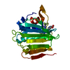
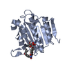
| Entry | Database: PDB / ID: 4em7 | ||||||
|---|---|---|---|---|---|---|---|
| Title | Crystal structure of a topoisomerase ATP inhibitor | ||||||
 Components Components | DNA topoisomerase IV, B subunit | ||||||
 Keywords Keywords | ISOMERASE/ISOMERASE INHIBITOR / protein-inhibitor complex / ATP binding / structure-based drug design / antimicrobial / virtual screen / ISOMERASE-ISOMERASE INHIBITOR complex | ||||||
| Function / homology | Histidine kinase-like ATPase, C-terminal domain / Heat Shock Protein 90 / 2-Layer Sandwich / Alpha Beta / Chem-0RA / :  Function and homology information Function and homology information | ||||||
| Biological species |  Streptococcus pneumoniae GA47373 (bacteria) Streptococcus pneumoniae GA47373 (bacteria) | ||||||
| Method |  X-RAY DIFFRACTION / SYNCHROTRON / MOLECULAR REPLACEMENT / molecular replacement / Resolution: 1.9 Å X-RAY DIFFRACTION / SYNCHROTRON / MOLECULAR REPLACEMENT / molecular replacement / Resolution: 1.9 Å | ||||||
 Authors Authors | Boriack-Sjodin, P.A. / Manchester, J. | ||||||
 Citation Citation | Journal: Bioorg.Med.Chem.Lett. / Year: 2012 Title: Discovery of a novel azaindole class of antibacterial agents targeting the ATPase domains of DNA gyrase and Topoisomerase IV. Authors: Manchester, J.I. / Dussault, D.D. / Rose, J.A. / Boriack-Sjodin, P.A. / Uria-Nickelsen, M. / Ioannidis, G. / Bist, S. / Fleming, P. / Hull, K.G. | ||||||
| History |
|
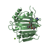
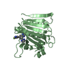
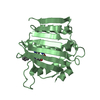
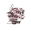
- Structure visualization
| Structure viewer | Molecule: MolmilJmol/JSmol |
|---|
- Downloads & links
-Download
| PDBx/mmCIF format | 4em7.cif.gz | 88.9 KB | Display | PDBx/mmCIF format |
|---|---|---|---|---|
| PDB format | pdb4em7.ent.gz | 67.9 KB | Display | PDB format |
| PDBx/mmJSON format | 4em7.json.gz | Tree view | PDBx/mmJSON format | |
| Others |  Other downloads Other downloads |
-Validation report
| Arichive directory | https://data.pdbj.org/pub/pdb/validation_reports/em/4em7ftp://data.pdbj.org/pub/pdb/validation_reports/em/4em7 | HTTPS FTP |
|---|
-Related structure data


-Links
 PDBj
PDBj- Assembly
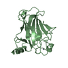
| Deposited unit | 
| ||||||||
|---|---|---|---|---|---|---|---|---|---|
| 1 |
| ||||||||
| Unit cell |
| ||||||||
| Components on special symmetry positions |
|
-Components
| #1: Protein | Mass: 24600.723 Da / Num. of mol.: 1 / Fragment: ATPase domain (UNP Residues 1-226) Source method: isolated from a genetically manipulated source Source: (gene. exp.) Streptococcus pneumoniae GA47373 (bacteria)Gene: ParE, SPAR94_0831 / Production host: References: UniProt: G6TGY9, Isomerases; Other isomerases; Sole sub-subclass for isomerases that do not belong in the other subclasses |
|---|---|
| #2: Chemical | ChemComp-0RA / 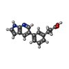  Mass: 266.295 Da / Num. of mol.: 1 / Source method: obtained synthetically / Formula: C16H14N2O2 Mass: 266.295 Da / Num. of mol.: 1 / Source method: obtained synthetically / Formula: C16H14N2O2 |
| #3: Water | ChemComp-HOH /  Mass: 18.015 Da / Num. of mol.: 112 / Source method: isolated from a natural source / Formula: H2O Mass: 18.015 Da / Num. of mol.: 112 / Source method: isolated from a natural source / Formula: H2O |
-Experimental details
-Experiment
| Experiment | Method: X-RAY DIFFRACTION / Number of used crystals: 1 |
|---|
- Sample preparation
| Crystal | Density Matthews: 2.16 Å3/Da / Density % sol: 43.07 % |
|---|---|
| Crystal grow | Temperature: 293 K / Method: inhibitor soak / pH: 7 Details: 18-25% Peg4000, 0.2M Ammonium Acetate, 0.1M MIB pH 7, inhibitor soak, temperature 293K |
-Data collection
| Diffraction | Mean temperature: 100 K | ||||||||||||||||||||||||||||||||||||||||||||||||||||||||||||||||||||||||||||||||||||||||
|---|---|---|---|---|---|---|---|---|---|---|---|---|---|---|---|---|---|---|---|---|---|---|---|---|---|---|---|---|---|---|---|---|---|---|---|---|---|---|---|---|---|---|---|---|---|---|---|---|---|---|---|---|---|---|---|---|---|---|---|---|---|---|---|---|---|---|---|---|---|---|---|---|---|---|---|---|---|---|---|---|---|---|---|---|---|---|---|---|---|
| Diffraction source | Source: SYNCHROTRON / Site: APS  / Beamline: 31-ID / Wavelength: 1 Å / Beamline: 31-ID / Wavelength: 1 Å | ||||||||||||||||||||||||||||||||||||||||||||||||||||||||||||||||||||||||||||||||||||||||
| Detector | Type: MAR CCD 165 mm / Detector: CCD / Date: Apr 3, 2008 | ||||||||||||||||||||||||||||||||||||||||||||||||||||||||||||||||||||||||||||||||||||||||
| Radiation | Protocol: SINGLE WAVELENGTH / Monochromatic (M) / Laue (L): M / Scattering type: x-ray | ||||||||||||||||||||||||||||||||||||||||||||||||||||||||||||||||||||||||||||||||||||||||
| Radiation wavelength | Wavelength: 1 Å / Relative weight: 1 | ||||||||||||||||||||||||||||||||||||||||||||||||||||||||||||||||||||||||||||||||||||||||
| Reflection | Resolution: 1.716→23.83 Å / Num. all: 17156 / Num. obs: 17156 / % possible obs: 99.9 % / Redundancy: 7.3 % / Rmerge(I) obs: 0.1 / Rsym value: 0.1 / Net I/σ(I): 15.3 | ||||||||||||||||||||||||||||||||||||||||||||||||||||||||||||||||||||||||||||||||||||||||
| Reflection shell | Diffraction-ID: 1
|
-Phasing
| Phasing | Method: molecular replacement |
|---|
- Processing
| Software |
| ||||||||||||||||||||||||||||||||||||||||||||||||||||||||||||
|---|---|---|---|---|---|---|---|---|---|---|---|---|---|---|---|---|---|---|---|---|---|---|---|---|---|---|---|---|---|---|---|---|---|---|---|---|---|---|---|---|---|---|---|---|---|---|---|---|---|---|---|---|---|---|---|---|---|---|---|---|---|
| Refinement | Method to determine structure: MOLECULAR REPLACEMENT / Resolution: 1.9→23.83 Å / Cor.coef. Fo:Fc: 0.95 / Cor.coef. Fo:Fc free: 0.935 / WRfactor Rfree: 0.2174 / WRfactor Rwork: 0.1888 / Occupancy max: 1 / Occupancy min: 0.5 / FOM work R set: 0.8766 / SU B: 5.697 / SU ML: 0.085 / SU R Cruickshank DPI: 0.1492 / SU Rfree: 0.1297 / Cross valid method: THROUGHOUT / σ(F): 0 / ESU R: 0.149 / ESU R Free: 0.13 / Stereochemistry target values: MAXIMUM LIKELIHOOD / Details: HYDROGENS HAVE BEEN ADDED IN THE RIDING POSITIONS
| ||||||||||||||||||||||||||||||||||||||||||||||||||||||||||||
| Solvent computation | Ion probe radii: 0.8 Å / Shrinkage radii: 0.8 Å / VDW probe radii: 1.2 Å / Solvent model: BABINET MODEL WITH MASK | ||||||||||||||||||||||||||||||||||||||||||||||||||||||||||||
| Displacement parameters | Biso max: 53.52 Å2 / Biso mean: 25.2291 Å2 / Biso min: 15.19 Å2
| ||||||||||||||||||||||||||||||||||||||||||||||||||||||||||||
| Refinement step | Cycle: LAST / Resolution: 1.9→23.83 Å
| ||||||||||||||||||||||||||||||||||||||||||||||||||||||||||||
| Refine LS restraints |
| ||||||||||||||||||||||||||||||||||||||||||||||||||||||||||||
| LS refinement shell | Resolution: 1.9→1.949 Å / Total num. of bins used: 20
| ||||||||||||||||||||||||||||||||||||||||||||||||||||||||||||
| Refinement TLS params. | Method: refined / Origin x: 23.266 Å / Origin y: 32.612 Å / Origin z: 0.553 Å
|
