 Movie
Movie Controller

[English] 日本語
 Yorodumi
- PDB-1ob5: T. aquaticus elongation factor EF-Tu complexed with the antibioti... -
+ Open data

- Basic information
| Entry | Database: PDB / ID: 1ob5 | ||||||
|---|---|---|---|---|---|---|---|
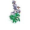
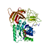
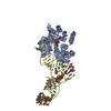
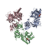
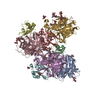
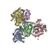
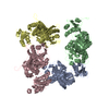
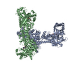
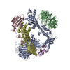
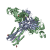
| Title | T. aquaticus elongation factor EF-Tu complexed with the antibiotic enacyloxin IIa, a GTP analog, and Phe-tRNA | ||||||
 Components Components |
| ||||||
 Keywords Keywords | HYDROLASE / GTPASE / TRANSLATION ELONGATION FACTOR / TRANSFER RNA / GTP-BINDING / NUCLEOTIDE-BINDING / PROTEIN BIOSYNTHESIS | ||||||
| Function / homology |  Function and homology information Function and homology informationprotein-synthesizing GTPase / translation elongation factor activity / GTPase activity / GTP binding / magnesium ion binding / cytosol Similarity search - Function | ||||||
| Biological species |   THERMUS AQUATICUS (bacteria) THERMUS AQUATICUS (bacteria) | ||||||
| Method |  X-RAY DIFFRACTION / SYNCHROTRON / MOLECULAR REPLACEMENT / Resolution: 3.1 Å X-RAY DIFFRACTION / SYNCHROTRON / MOLECULAR REPLACEMENT / Resolution: 3.1 Å | ||||||
 Authors Authors | Dahlberg, C. / Nielsen, R.C. / Parmeggiani, A. / Nyborg, J. / Nissen, P. | ||||||
 Citation Citation | Journal: J.Biol.Chem. / Year: 2006 Title: Enacyloxin Iia Pinpoints a Binding Pocket of Elongation Factor TU for Development of Novel Antibiotics. Authors: Parmeggiani, A. / Krab, I.M. / Watanabe, T. / Nielsen, R.C. / Dahlberg, C. / Nyborg, J. / Nissen, P. | ||||||
| History |
| ||||||
| Remark 700 | SHEET THE SHEET STRUCTURE OF THIS MOLECULE IS BIFURCATED. IN ORDER TO REPRESENT THIS FEATURE IN ... SHEET THE SHEET STRUCTURE OF THIS MOLECULE IS BIFURCATED. IN ORDER TO REPRESENT THIS FEATURE IN THE SHEET RECORDS BELOW, TWO SHEETS ARE DEFINED. |
- Structure visualization
| Structure viewer | Molecule: MolmilJmol/JSmol |
|---|
- Downloads & links
-Download
| PDBx/mmCIF format | 1ob5.cif.gz | 387.1 KB | Display | PDBx/mmCIF format |
|---|---|---|---|---|
| PDB format | pdb1ob5.ent.gz | 306.8 KB | Display | PDB format |
| PDBx/mmJSON format | 1ob5.json.gz | Tree view | PDBx/mmJSON format | |
| Others |  Other downloads Other downloads |
-Validation report
| Arichive directory | https://data.pdbj.org/pub/pdb/validation_reports/ob/1ob5ftp://data.pdbj.org/pub/pdb/validation_reports/ob/1ob5 | HTTPS FTP |
|---|
-Related structure data
| Related structure data | 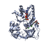 2bvnC  1ld1 S: Starting model for refinement C: citing same article ( |
|---|---|
| Similar structure data |


-Links
 PDBj
PDBj


- Assembly
| Deposited unit | 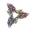
| ||||||||
|---|---|---|---|---|---|---|---|---|---|
| 1 |
| ||||||||
| Unit cell |
|
-Components
| #1: Protein | Mass: 44742.980 Da / Num. of mol.: 3 Source method: isolated from a genetically manipulated source Source: (gene. exp.) THERMUS AQUATICUS (bacteria) / Description: TUF-A GENE / Production host: #2: RNA chain | Mass: 25326.479 Da / Num. of mol.: 3 / Source method: isolated from a natural source / Details: SIGMA-ALDRICH COMPOUND / Source: (natural) #3: Chemical |   Mass: 522.196 Da / Num. of mol.: 3 / Source method: obtained synthetically / Formula: C10H17N6O13P3 Mass: 522.196 Da / Num. of mol.: 3 / Source method: obtained synthetically / Formula: C10H17N6O13P3Comment: GppNHp, GMPPNP, energy-carrying molecule analogue*YM #4: Chemical |   Mass: 24.305 Da / Num. of mol.: 3 / Source method: obtained synthetically / Formula: Mg Mass: 24.305 Da / Num. of mol.: 3 / Source method: obtained synthetically / Formula: Mg#5: Chemical |   Mass: 702.616 Da / Num. of mol.: 3 / Source method: obtained synthetically / Formula: C33H45Cl2NO11 Mass: 702.616 Da / Num. of mol.: 3 / Source method: obtained synthetically / Formula: C33H45Cl2NO11Compound details | PROMOTES THE GTP-DEPENDENT BINDING OF AMINOACYL-TRNA TO THE A-SITE OF RIBOSOMES DURING PROTEIN BIOSYNTHES | |
|---|
-Experimental details
-Experiment
| Experiment | Method: X-RAY DIFFRACTION / Number of used crystals: 1 |
|---|
- Sample preparation
| Crystal | Density Matthews: 3.6 Å3/Da / Density % sol: 64 % |
|---|---|
| Crystal grow | pH: 6.8 Details: 1.8M AMMONIUM SULPHATE, 10 MM MAGNESIUM CHLORIDE,20 MM TRIS-MES, PH 6.4 |
-Data collection
| Diffraction | Mean temperature: 100 K |
|---|---|
| Diffraction source | Source: SYNCHROTRON / Site: EMBL/DESY, HAMBURG  / Beamline: X13 / Wavelength: 0.802 / Beamline: X13 / Wavelength: 0.802 |
| Detector | Type: MARRESEARCH / Detector: CCD / Date: Apr 4, 2002 |
| Radiation | Protocol: SINGLE WAVELENGTH / Monochromatic (M) / Laue (L): M / Scattering type: x-ray |
| Radiation wavelength | Wavelength: 0.802 Å / Relative weight: 1 |
| Reflection | Resolution: 3.33→30 Å / Num. obs: 48311 / % possible obs: 99.9 % / Observed criterion σ(I): -3 / Redundancy: 6.3 % / Biso Wilson estimate: 68.3 Å2 / Rmerge(I) obs: 0.12 / Net I/σ(I): 12 |
| Reflection shell | Resolution: 3.3→3.38 Å / Redundancy: 5.7 % / Rmerge(I) obs: 0.518 / Mean I/σ(I) obs: 4.6 / % possible all: 99.9 |
- Processing
| Software |
| ||||||||||||||||||||||||||||||||||||||||||||||||||||||||||||||||||||||||||||||||
|---|---|---|---|---|---|---|---|---|---|---|---|---|---|---|---|---|---|---|---|---|---|---|---|---|---|---|---|---|---|---|---|---|---|---|---|---|---|---|---|---|---|---|---|---|---|---|---|---|---|---|---|---|---|---|---|---|---|---|---|---|---|---|---|---|---|---|---|---|---|---|---|---|---|---|---|---|---|---|---|---|---|
| Refinement | Method to determine structure: MOLECULAR REPLACEMENT Starting model: PDB ENTRY 1LD1 1ld1 Resolution: 3.1→29.54 Å / Rfactor Rfree error: 0.008 / Data cutoff high absF: 8253666.21 / Data cutoff low absF: 0 / Cross valid method: THROUGHOUT / σ(F): 0
| ||||||||||||||||||||||||||||||||||||||||||||||||||||||||||||||||||||||||||||||||
| Solvent computation | Solvent model: FLAT MODEL / Bsol: 59.7579 Å2 / ksol: 0.290638 e/Å3 | ||||||||||||||||||||||||||||||||||||||||||||||||||||||||||||||||||||||||||||||||
| Displacement parameters | Biso mean: 76.5 Å2
| ||||||||||||||||||||||||||||||||||||||||||||||||||||||||||||||||||||||||||||||||
| Refine analyze |
| ||||||||||||||||||||||||||||||||||||||||||||||||||||||||||||||||||||||||||||||||
| Refinement step | Cycle: LAST / Resolution: 3.1→29.54 Å
| ||||||||||||||||||||||||||||||||||||||||||||||||||||||||||||||||||||||||||||||||
| Refine LS restraints |
| ||||||||||||||||||||||||||||||||||||||||||||||||||||||||||||||||||||||||||||||||
| Xplor file |
|
