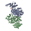
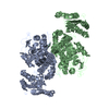
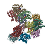
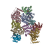
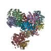
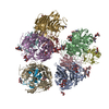
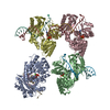
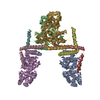
Entry Database : PDB / ID : 5elpTitle Ketosynthase from module 1 of the bacillaene synthase from Bacillus amyloliquefaciens FZB42 NRPS/PKS protein Keywords / / / / Function / homology Function Domain/homology Component
/ / / / / / / / / / / / / / / / / / / / / / / / / / / / / / / / / / / / / / / / / / / / / / / / / / / / / / / / / / / / / / / / / / / / / Biological species Bacillus amyloliquefaciens (bacteria)Method / / Resolution : 2.93 Å Authors Wagner, D.T. / Gay, D.C. / Keatinge-Clay, A.T. / Zogzas, C.E. Journal : J.Struct.Biol. / Year : 2016Title : The LINKS motif zippers trans-acyltransferase polyketide synthase assembly lines into a biosynthetic megacomplex.Authors : Gay, D.C. / Wagner, D.T. / Meinke, J.L. / Zogzas, C.E. / Gay, G.R. / Keatinge-Clay, A.T. History Deposition Nov 4, 2015 Deposition site / Processing site Revision 1.0 Jan 13, 2016 Provider / Type Revision 1.1 Nov 2, 2016 Group Revision 1.2 Nov 13, 2024 Group Data collection / Database references ... Data collection / Database references / Derived calculations / Refinement description / Structure summary Category chem_comp_atom / chem_comp_bond ... chem_comp_atom / chem_comp_bond / citation / database_2 / pdbx_entry_details / pdbx_modification_feature / pdbx_struct_oper_list / struct_conn / struct_ncs_dom_lim Item _citation.journal_id_CSD / _database_2.pdbx_DOI ... _citation.journal_id_CSD / _database_2.pdbx_DOI / _database_2.pdbx_database_accession / _pdbx_struct_oper_list.symmetry_operation / _struct_conn.pdbx_dist_value / _struct_conn.ptnr1_auth_asym_id / _struct_conn.ptnr1_auth_comp_id / _struct_conn.ptnr1_auth_seq_id / _struct_conn.ptnr1_label_asym_id / _struct_conn.ptnr1_label_atom_id / _struct_conn.ptnr1_label_comp_id / _struct_conn.ptnr1_label_seq_id / _struct_conn.ptnr2_auth_asym_id / _struct_conn.ptnr2_auth_comp_id / _struct_conn.ptnr2_auth_seq_id / _struct_conn.ptnr2_label_asym_id / _struct_conn.ptnr2_label_atom_id / _struct_conn.ptnr2_label_comp_id / _struct_conn.ptnr2_label_seq_id / _struct_conn.ptnr2_symmetry / _struct_ncs_dom_lim.beg_auth_comp_id / _struct_ncs_dom_lim.beg_label_asym_id / _struct_ncs_dom_lim.beg_label_comp_id / _struct_ncs_dom_lim.beg_label_seq_id / _struct_ncs_dom_lim.end_auth_comp_id / _struct_ncs_dom_lim.end_label_asym_id / _struct_ncs_dom_lim.end_label_comp_id / _struct_ncs_dom_lim.end_label_seq_id
Show all Show less
 Movie
Movie Controller

 Yorodumi
 Open data
 Basic information
 Components
 Keywords
 Function and homology information
 X-RAY DIFFRACTION /
  Authors
 Citation
 Structure visualization
 Downloads & links
 Other downloads


 PDBj
PDBj


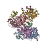
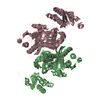
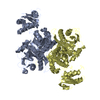
 Assembly
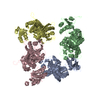
 Sample preparation
 / Beamline: 23-ID-D / Wavelength: 1 Å
 Processing