 Movie
Movie Controller

[English] 日本語
 Yorodumi
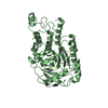
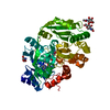
- PDB-1o5h: Crystal structure of formiminotetrahydrofolate cyclodeaminase (TM... -
+ Open data

- Basic information
| Entry | Database: PDB / ID: 1o5h | ||||||
|---|---|---|---|---|---|---|---|
| Title | Crystal structure of formiminotetrahydrofolate cyclodeaminase (TM1560) from Thermotoga maritima at 2.80 A resolution | ||||||
 Components Components | Formiminotetrahydrofolate cyclodeaminase | ||||||
 Keywords Keywords | LYASE / TM1560 / Formiminotetrahydrofolate cyclodeaminase / STRUCTURAL GENOMICS / JCSG / PSI / Protein Structure Initiative / Joint Center for Structural Genomics | ||||||
| Function / homology | Formiminotetrahydrofolate cyclodeaminase monomer, up-and-down helical bundle / Cyclodeaminase/cyclohydrolase / Formimidoyltransferase-cyclodeaminase-like superfamily / Formiminotransferase-cyclodeaminase / catalytic activity / Four Helix Bundle (Hemerythrin (Met), subunit A) / Up-down Bundle / Mainly Alpha / Serine cycle enzyme, putative Function and homology information Function and homology information | ||||||
| Biological species |   Thermotoga maritima (bacteria) Thermotoga maritima (bacteria) | ||||||
| Method |  X-RAY DIFFRACTION / SYNCHROTRON / SAD / Resolution: 2.8 Å X-RAY DIFFRACTION / SYNCHROTRON / SAD / Resolution: 2.8 Å | ||||||
 Authors Authors | Joint Center for Structural Genomics (JCSG) | ||||||
 Citation Citation | Journal: Proteins / Year: 2005 Title: Crystal structure of a formiminotetrahydrofolate cyclodeaminase (TM1560) from Thermotoga maritima at 2.80 A resolution reveals a new fold Authors: Xu, Q. / Schwarzenbacher, R. / McMullan, D. / Abdubek, P. / Ambing, E. / Biorac, T. / Canaves, J.M. / Chiu, H.J. / Dai, X. / Deacon, A.M. / DiDonato, M. / Elsliger, M.A. / Godzik, A. / ...Authors: Xu, Q. / Schwarzenbacher, R. / McMullan, D. / Abdubek, P. / Ambing, E. / Biorac, T. / Canaves, J.M. / Chiu, H.J. / Dai, X. / Deacon, A.M. / DiDonato, M. / Elsliger, M.A. / Godzik, A. / Grittini, C. / Grzechnik, S.K. / Hampton, E. / Hornsby, M. / Jaroszewski, L. / Klock, H.E. / Koesema, E. / Kreusch, A. / Kuhn, P. / Lesley, S.A. / Levin, I. / Miller, M.D. / Morse, A. / Moy, K. / Ouyang, J. / Page, R. / Quijano, K. / Reyes, R. / Robb, A. / Sims, E. / Spraggon, G. / Stevens, R.C. / van den Bedem, H. / Velasquez, J. / Vincent, J. / von Delft, F. / Wang, X. / West, B. / White, A. / Wolf, G. / Zagnitko, O. / Hodgson, K.O. / Wooley, J. / Wilson, I.A. | ||||||
| History |
|
- Structure visualization
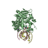
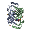
| Structure viewer | Molecule: MolmilJmol/JSmol |
|---|
- Downloads & links
-Download
| PDBx/mmCIF format | 1o5h.cif.gz | 88.7 KB | Display | PDBx/mmCIF format |
|---|---|---|---|---|
| PDB format | pdb1o5h.ent.gz | 69.3 KB | Display | PDB format |
| PDBx/mmJSON format | 1o5h.json.gz | Tree view | PDBx/mmJSON format | |
| Others |  Other downloads Other downloads |
-Validation report
| Arichive directory | https://data.pdbj.org/pub/pdb/validation_reports/o5/1o5hftp://data.pdbj.org/pub/pdb/validation_reports/o5/1o5h | HTTPS FTP |
|---|
-Related structure data
| Similar structure data | |
|---|---|
| Other databases |


-Links
 PDBj
PDBj- Assembly
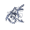
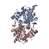
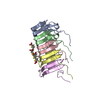
| Deposited unit | 
| ||||||||||||||||||||||||||||||||||||||||||||||||||||||||||||||||||||||||||||||||||||||||||||||||||||||||||||||||||||||||||||||||||||||||||||||||||||||||||
|---|---|---|---|---|---|---|---|---|---|---|---|---|---|---|---|---|---|---|---|---|---|---|---|---|---|---|---|---|---|---|---|---|---|---|---|---|---|---|---|---|---|---|---|---|---|---|---|---|---|---|---|---|---|---|---|---|---|---|---|---|---|---|---|---|---|---|---|---|---|---|---|---|---|---|---|---|---|---|---|---|---|---|---|---|---|---|---|---|---|---|---|---|---|---|---|---|---|---|---|---|---|---|---|---|---|---|---|---|---|---|---|---|---|---|---|---|---|---|---|---|---|---|---|---|---|---|---|---|---|---|---|---|---|---|---|---|---|---|---|---|---|---|---|---|---|---|---|---|---|---|---|---|---|---|---|
| 1 |
| ||||||||||||||||||||||||||||||||||||||||||||||||||||||||||||||||||||||||||||||||||||||||||||||||||||||||||||||||||||||||||||||||||||||||||||||||||||||||||
| Unit cell |
| ||||||||||||||||||||||||||||||||||||||||||||||||||||||||||||||||||||||||||||||||||||||||||||||||||||||||||||||||||||||||||||||||||||||||||||||||||||||||||
| Noncrystallographic symmetry (NCS) | NCS domain:
NCS domain segments: Ens-ID: 1
|
-Components
| #1: Protein | Mass: 24815.568 Da / Num. of mol.: 2 Source method: isolated from a genetically manipulated source Source: (gene. exp.) Thermotoga maritima (bacteria) / Gene: TM1560 / Production host: References: UniProt: Q9X1P6, formimidoyltetrahydrofolate cyclodeaminase Has protein modification | Y | |
|---|
-Experimental details
-Experiment
| Experiment | Method: X-RAY DIFFRACTION / Number of used crystals: 1 |
|---|
- Sample preparation
| Crystal | Density Matthews: 2.84 Å3/Da / Density % sol: 56.4 % | |||||||||||||||||||||||||||||||||||
|---|---|---|---|---|---|---|---|---|---|---|---|---|---|---|---|---|---|---|---|---|---|---|---|---|---|---|---|---|---|---|---|---|---|---|---|---|
| Crystal grow | Temperature: 277 K / Method: vapor diffusion, sitting drop, nanodrop / pH: 5 Details: 5% PEG-6000, 0.1M citric acid pH 5.0, VAPOR DIFFUSION,SITTING DROP,NANODROP, temperature 277K | |||||||||||||||||||||||||||||||||||
| Crystal grow | *PLUS pH: 7.9 / Method: vapor diffusion | |||||||||||||||||||||||||||||||||||
| Components of the solutions | *PLUS
|
-Data collection
| Diffraction | Mean temperature: 100 K |
|---|---|
| Diffraction source | Source: SYNCHROTRON / Site: SSRL  / Beamline: BL9-1 / Wavelength: 0.978998 / Beamline: BL9-1 / Wavelength: 0.978998 |
| Detector | Type: ADSC QUANTUM 315 / Detector: CCD / Date: Nov 21, 2002 |
| Radiation | Monochromator: single crystal Si(311) bent monochromator / Protocol: SINGLE WAVELENGTH / Monochromatic (M) / Laue (L): M / Scattering type: x-ray |
| Radiation wavelength | Wavelength: 0.978998 Å / Relative weight: 1 |
| Reflection | Resolution: 2.6→42 Å / Num. all: 13727 / Num. obs: 13727 / % possible obs: 88.3 % / Redundancy: 6.4 % / Biso Wilson estimate: 64.28 Å2 / Rsym value: 0.108 / Net I/σ(I): 13.3 |
| Reflection shell | Resolution: 2.6→2.67 Å / Redundancy: 3.2 % / Mean I/σ(I) obs: 1.4 / Num. unique all: 419 / Rsym value: 0.591 / % possible all: 38.5 |
| Reflection | *PLUS Highest resolution: 2.8 Å / Num. obs: 12284 / % possible obs: 97.1 % / Num. measured all: 81783 / Rmerge(I) obs: 0.067 |
| Reflection shell | *PLUS Highest resolution: 2.8 Å / Lowest resolution: 2.87 Å / % possible obs: 76.5 % / Rmerge(I) obs: 0.47 |
- Processing
| Software |
| |||||||||||||||||||||||||||||||||||||||||||||||||||||||||||||||||||||||||||||||||||||
|---|---|---|---|---|---|---|---|---|---|---|---|---|---|---|---|---|---|---|---|---|---|---|---|---|---|---|---|---|---|---|---|---|---|---|---|---|---|---|---|---|---|---|---|---|---|---|---|---|---|---|---|---|---|---|---|---|---|---|---|---|---|---|---|---|---|---|---|---|---|---|---|---|---|---|---|---|---|---|---|---|---|---|---|---|---|---|
| Refinement | Method to determine structure: SAD / Resolution: 2.8→42 Å / Cor.coef. Fo:Fc: 0.949 / Cor.coef. Fo:Fc free: 0.891 / SU B: 30.538 / SU ML: 0.273 / TLS residual ADP flag: LIKELY RESIDUAL / Isotropic thermal model: ISOTROPIC / Cross valid method: THROUGHOUT / ESU R Free: 0.411 Stereochemistry target values: MAXIMUM LIKELIHOOD WITH PHASES Details: 1. THE CONFORMATION OF RESIDUES 20-22 (P-T-P) IS SUSPECT, BEING POORLY DEFINED IN EXPERIMENTAL MAPS AND WITH RESIDUAL DIFFERENCE DENSITY IN THE REFINED STRUCTURE, AS WELL AS UNUSUAL PRO ...Details: 1. THE CONFORMATION OF RESIDUES 20-22 (P-T-P) IS SUSPECT, BEING POORLY DEFINED IN EXPERIMENTAL MAPS AND WITH RESIDUAL DIFFERENCE DENSITY IN THE REFINED STRUCTURE, AS WELL AS UNUSUAL PRO PUCKERING PHASES AFTER 2. RESIDUES 88-98 IN SUBUNIT ARE POORLY ORDERED IN SUBUNIT A AND DEVIATE CONSIDERABLY FROM SUBUNIT B; NCS RESTRAINTS WERE KEPT TIGHT HERE, HOWEVER, SINCE LOOSE RESTRAINTS DID NOT IMPROVE RFREE OR THE GEOMETRY. 3. THE TWO C-TERMINAL RESIDUES OF SUBUNIT B WERE DISORDERED OMITTED IN THE MODEL.
| |||||||||||||||||||||||||||||||||||||||||||||||||||||||||||||||||||||||||||||||||||||
| Solvent computation | Ion probe radii: 0.8 Å / Shrinkage radii: 0.8 Å / VDW probe radii: 1.2 Å / Solvent model: BABINET MODEL WITH MASK | |||||||||||||||||||||||||||||||||||||||||||||||||||||||||||||||||||||||||||||||||||||
| Displacement parameters | Biso mean: 44.593 Å2
| |||||||||||||||||||||||||||||||||||||||||||||||||||||||||||||||||||||||||||||||||||||
| Refinement step | Cycle: LAST / Resolution: 2.8→42 Å
| |||||||||||||||||||||||||||||||||||||||||||||||||||||||||||||||||||||||||||||||||||||
| Refine LS restraints |
| |||||||||||||||||||||||||||||||||||||||||||||||||||||||||||||||||||||||||||||||||||||
| Refine LS restraints NCS | Dom-ID: 1 / Auth asym-ID: A / Ens-ID: 1 / Refine-ID: X-RAY DIFFRACTION
| |||||||||||||||||||||||||||||||||||||||||||||||||||||||||||||||||||||||||||||||||||||
| LS refinement shell | Resolution: 2.8→2.873 Å / Total num. of bins used: 20
| |||||||||||||||||||||||||||||||||||||||||||||||||||||||||||||||||||||||||||||||||||||
| Refinement TLS params. | Method: refined / Refine-ID: X-RAY DIFFRACTION
| |||||||||||||||||||||||||||||||||||||||||||||||||||||||||||||||||||||||||||||||||||||
| Refinement TLS group | Refine-ID: X-RAY DIFFRACTION / Selection: ALL
| |||||||||||||||||||||||||||||||||||||||||||||||||||||||||||||||||||||||||||||||||||||
| Refinement | *PLUS Num. reflection obs: 12173 / Rfactor Rfree: 0.278 / Rfactor Rwork: 0.199 | |||||||||||||||||||||||||||||||||||||||||||||||||||||||||||||||||||||||||||||||||||||
| Solvent computation | *PLUS | |||||||||||||||||||||||||||||||||||||||||||||||||||||||||||||||||||||||||||||||||||||
| Displacement parameters | *PLUS | |||||||||||||||||||||||||||||||||||||||||||||||||||||||||||||||||||||||||||||||||||||
| Refine LS restraints | *PLUS
|
