 Movie
Movie Controller

[English] 日本語
 Yorodumi
- PDB-1m7j: Crystal structure of D-aminoacylase defines a novel subset of ami... -
+ Open data

- Basic information
| Entry | Database: PDB / ID: 1m7j | ||||||
|---|---|---|---|---|---|---|---|
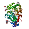
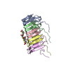
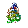
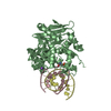
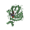
| Title | Crystal structure of D-aminoacylase defines a novel subset of amidohydrolases | ||||||
 Components Components | D-aminoacylase | ||||||
 Keywords Keywords | HYDROLASE / TIN-barrel / metal-dependent amidohydrolase | ||||||
| Function / homology |  Function and homology information Function and homology informationN-acyl-D-amino-acid deacylase / N-acyl-D-amino-acid deacylase activity / hydrolase activity, acting on carbon-nitrogen (but not peptide) bonds, in cyclic amides / metal ion binding / cytosol Similarity search - Function | ||||||
| Biological species |  Alcaligenes faecalis (bacteria) Alcaligenes faecalis (bacteria) | ||||||
| Method |  X-RAY DIFFRACTION / SYNCHROTRON / SAD / Resolution: 1.5 Å X-RAY DIFFRACTION / SYNCHROTRON / SAD / Resolution: 1.5 Å | ||||||
 Authors Authors | Liaw, S.-H. / Chen, S.-J. / Ko, T.-P. / Hsu, C.-S. / Wang, A.H.-J. / Tsai, Y.-C. | ||||||
 Citation Citation | Journal: J.Biol.Chem. / Year: 2003 Title: Crystal Structure of D-Aminoacylase from Alcaligenes faecalis DA1. A NOVEL SUBSET OF AMIDOHYDROLASES AND INSIGHTS INTO THE ENZYME MECHANISM. Authors: Liaw, S.-H. / Chen, S.-J. / Ko, T.-P. / Hsu, C.-S. / Chen, C.J. / Wang, A.H. / Tsai, Y.-C. #1: Journal: Protein Sci. / Year: 2002Title: Structural-based mutational analysis of D-aminoacylase from Alcaligenes faecalis DA1. Authors: Hsu, C.-S. / Lai, W.-L. / Chang, W.-W. / Liaw, S.-H. / Tsai, Y.-C. #2: Journal: Acta Crystallogr.,Sect.D / Year: 2002Title: Crystallization and preliminary crystallographic analysis of a D-aminoacylase from Alcaligenes faecalis DA1 Authors: Hsu, C.-S. / Chen, S.-J. / Tsai, Y.-C. / Lin, T.-W. / Liaw, S.-H. / Wang, A.H. | ||||||
| History |
|
- Structure visualization
| Structure viewer | Molecule: MolmilJmol/JSmol |
|---|
- Downloads & links
-Download
| PDBx/mmCIF format | 1m7j.cif.gz | 114.3 KB | Display | PDBx/mmCIF format |
|---|---|---|---|---|
| PDB format | pdb1m7j.ent.gz | 85.8 KB | Display | PDB format |
| PDBx/mmJSON format | 1m7j.json.gz | Tree view | PDBx/mmJSON format | |
| Others |  Other downloads Other downloads |
-Validation report
| Arichive directory | https://data.pdbj.org/pub/pdb/validation_reports/m7/1m7jftp://data.pdbj.org/pub/pdb/validation_reports/m7/1m7j | HTTPS FTP |
|---|
-Related structure data
| Similar structure data |
|---|


-Links
 PDBj
PDBj

- Assembly
| Deposited unit | 
| ||||||||
|---|---|---|---|---|---|---|---|---|---|
| 1 |
| ||||||||
| Unit cell |
| ||||||||
| Details | This protein is monomeric. |
-Components
| #1: Protein | Mass: 52060.707 Da / Num. of mol.: 1 Source method: isolated from a genetically manipulated source Source: (gene. exp.) Alcaligenes faecalis (bacteria) / Plasmid: pQE / Production host: | ||||
|---|---|---|---|---|---|
| #2: Chemical |   Mass: 65.409 Da / Num. of mol.: 2 / Source method: obtained synthetically / Formula: Zn Mass: 65.409 Da / Num. of mol.: 2 / Source method: obtained synthetically / Formula: Zn#3: Chemical |   Mass: 59.044 Da / Num. of mol.: 2 / Source method: obtained synthetically / Formula: C2H3O2 Mass: 59.044 Da / Num. of mol.: 2 / Source method: obtained synthetically / Formula: C2H3O2#4: Water | ChemComp-HOH / |  Mass: 18.015 Da / Num. of mol.: 503 / Source method: isolated from a natural source / Formula: H2O Mass: 18.015 Da / Num. of mol.: 503 / Source method: isolated from a natural source / Formula: H2O |
-Experimental details
-Experiment
| Experiment | Method: X-RAY DIFFRACTION / Number of used crystals: 1 |
|---|
- Sample preparation
| Crystal | Density Matthews: 2.74 Å3/Da / Density % sol: 54.79 % | ||||||||||||||||||||||||||||||
|---|---|---|---|---|---|---|---|---|---|---|---|---|---|---|---|---|---|---|---|---|---|---|---|---|---|---|---|---|---|---|---|
| Crystal grow | Temperature: 295 K / Method: vapor diffusion, hanging drop / pH: 5.6 Details: sodium citrate, ammonium acetate, PEG 4000, pH 5.6, VAPOR DIFFUSION, HANGING DROP, temperature 295K | ||||||||||||||||||||||||||||||
| Crystal grow | *PLUS Details: Hsu, C.-S., (2002) Acta Crystallogr., Sect.D, 58, 1482. | ||||||||||||||||||||||||||||||
| Components of the solutions | *PLUS
|
-Data collection
| Diffraction |
| |||||||||||||||
|---|---|---|---|---|---|---|---|---|---|---|---|---|---|---|---|---|
| Diffraction source |
| |||||||||||||||
| Detector |
| |||||||||||||||
| Radiation | Monochromator: rotated-inclined double-crystal monochromator Protocol: SINGLE WAVELENGTH / Monochromatic (M) / Laue (L): M / Scattering type: x-ray | |||||||||||||||
| Radiation wavelength |
| |||||||||||||||
| Reflection | Resolution: 1.5→50 Å / Num. all: 101683 / Num. obs: 100783 / % possible obs: 99.2 % / Observed criterion σ(F): 1 / Observed criterion σ(I): 1 / Redundancy: 6.3 % / Rmerge(I) obs: 0.047 / Net I/σ(I): 50.5 | |||||||||||||||
| Reflection shell | Resolution: 1.5→1.55 Å / Redundancy: 6.6 % / Rmerge(I) obs: 0.116 / Mean I/σ(I) obs: 17.6 / Num. unique all: 9984 / % possible all: 100 | |||||||||||||||
| Reflection | *PLUS Lowest resolution: 150 Å | |||||||||||||||
| Reflection shell | *PLUS Highest resolution: 1.5 Å / % possible obs: 100 % / Num. unique obs: 9984 |

- Processing
| Software |
| |||||||||||||||||||||||||
|---|---|---|---|---|---|---|---|---|---|---|---|---|---|---|---|---|---|---|---|---|---|---|---|---|---|---|
| Refinement | Method to determine structure: SAD / Resolution: 1.5→50 Å / Cross valid method: THROUGHOUT / σ(F): 0 / σ(I): 0 / Stereochemistry target values: Engh & Huber
| |||||||||||||||||||||||||
| Displacement parameters | Biso mean: 10.8 Å2 | |||||||||||||||||||||||||
| Refine analyze |
| |||||||||||||||||||||||||
| Refinement step | Cycle: LAST / Resolution: 1.5→50 Å
| |||||||||||||||||||||||||
| Refine LS restraints |
| |||||||||||||||||||||||||
| LS refinement shell | Resolution: 1.5→1.55 Å
| |||||||||||||||||||||||||
| Refinement | *PLUS Lowest resolution: 150 Å / Rfactor Rwork: 0.16 | |||||||||||||||||||||||||
| Solvent computation | *PLUS | |||||||||||||||||||||||||
| Displacement parameters | *PLUS |
