 Movie
Movie Controller

[English] 日本語
 Yorodumi
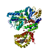
- PDB-1o4x: TERNARY COMPLEX OF THE DNA BINDING DOMAINS OF THE OCT1 AND SOX2 T... -
+ Open data

- Basic information
| Entry | Database: PDB / ID: 1o4x | ||||||
|---|---|---|---|---|---|---|---|
| Title | TERNARY COMPLEX OF THE DNA BINDING DOMAINS OF THE OCT1 AND SOX2 TRANSCRIPTION FACTORS WITH A 19MER OLIGONUCLEOTIDE FROM THE HOXB1 REGULATORY ELEMENT | ||||||
 Components Components |
| ||||||
 Keywords Keywords | TRANSCRIPTION/DNA / OCT1 / POU / POUS / POUHD / SOX2 / HMG-BOX / TRANSCRIPTION FACTORS / DNA / PROTEIN-DNA COMPLEX / TRANSCRIPTION-DNA COMPLEX | ||||||
| Function / homology |  Function and homology information Function and homology informationglial cell fate commitment / regulation of myofibroblast cell apoptotic process / Formation of the posterior neural plate / POU5F1 (OCT4), SOX2, NANOG repress genes related to differentiation / Formation of the anterior neural plate / endodermal cell fate specification / response to oxygen-glucose deprivation / adenohypophysis development / negative regulation of cell cycle G1/S phase transition / POU5F1 (OCT4), SOX2, NANOG activate genes related to proliferation ...glial cell fate commitment / regulation of myofibroblast cell apoptotic process / Formation of the posterior neural plate / POU5F1 (OCT4), SOX2, NANOG repress genes related to differentiation / Formation of the anterior neural plate / endodermal cell fate specification / response to oxygen-glucose deprivation / adenohypophysis development / negative regulation of cell cycle G1/S phase transition / POU5F1 (OCT4), SOX2, NANOG activate genes related to proliferation / Specification of the neural plate border / pituitary gland development / Regulation of MITF-M-dependent genes involved in extracellular matrix, focal adhesion and epithelial-to-mesenchymal transition / positive regulation of cell-cell adhesion / Transcriptional regulation of pluripotent stem cells / Transcriptional Regulation by MECP2 / neuronal stem cell population maintenance / Germ layer formation at gastrulation / eye development / tissue regeneration / response to growth factor / RNA Polymerase III Transcription Initiation From Type 3 Promoter / RNA Polymerase III Abortive And Retractive Initiation / miRNA binding / negative regulation of neuron differentiation / forebrain development / inner ear development / nitric-oxide synthase binding / RNA polymerase II transcribes snRNA genes / somatic stem cell population maintenance / RNA polymerase II core promoter sequence-specific DNA binding / Transcriptional and post-translational regulation of MITF-M expression and activity / positive regulation of cell differentiation / Deactivation of the beta-catenin transactivating complex / negative regulation of canonical Wnt signaling pathway / brain development / positive regulation of miRNA transcription / response to wounding / RNA polymerase II transcription regulator complex / neuron differentiation / osteoblast differentiation / chromatin organization / regulation of gene expression / Interleukin-4 and Interleukin-13 signaling / DNA-binding transcription activator activity, RNA polymerase II-specific / transcription regulator complex / cellular response to hypoxia / Estrogen-dependent gene expression / sequence-specific DNA binding / response to ethanol / DNA-binding transcription factor activity, RNA polymerase II-specific / positive regulation of MAPK cascade / transcription cis-regulatory region binding / nuclear speck / RNA polymerase II cis-regulatory region sequence-specific DNA binding / DNA-binding transcription factor activity / negative regulation of DNA-templated transcription / regulation of transcription by RNA polymerase II / regulation of DNA-templated transcription / positive regulation of DNA-templated transcription / chromatin / negative regulation of transcription by RNA polymerase II / endoplasmic reticulum / positive regulation of transcription by RNA polymerase II / DNA binding / nucleoplasm / identical protein binding / nucleus / cytoplasm / cytosol Similarity search - Function | ||||||
| Biological species |  Homo sapiens (human) Homo sapiens (human) | ||||||
| Method | SOLUTION NMR / CONJOINED RIGID BODY, TORSION ANGLE DYNAMICS | ||||||
 Authors Authors | Clore, G.M. / Williams, D.C. | ||||||
 Citation Citation | Journal: J.Biol.Chem. / Year: 2004 Title: Molecular basis for synergistic transcriptional activation by Oct1 and Sox2 revealed from the solution structure of the 42-kDa Oct1.Sox2.Hoxb1-DNA ternary transcription factor complex. Authors: Williams, D.C. / Cai, M. / Clore, G.M. | ||||||
| History |
|
- Structure visualization
| Structure viewer | Molecule: MolmilJmol/JSmol |
|---|
- Downloads & links
-Download
| PDBx/mmCIF format | 1o4x.cif.gz | 118.3 KB | Display | PDBx/mmCIF format |
|---|---|---|---|---|
| PDB format | pdb1o4x.ent.gz | 90 KB | Display | PDB format |
| PDBx/mmJSON format | 1o4x.json.gz | Tree view | PDBx/mmJSON format | |
| Others |  Other downloads Other downloads |
-Validation report
| Arichive directory | https://data.pdbj.org/pub/pdb/validation_reports/o4/1o4xftp://data.pdbj.org/pub/pdb/validation_reports/o4/1o4x | HTTPS FTP |
|---|
-Related structure data
| Similar structure data |
|---|


-Links
 PDBj
PDBj


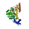
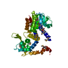
- Assembly
| Deposited unit | 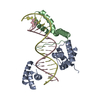
| |||||||||
|---|---|---|---|---|---|---|---|---|---|---|
| 1 |
| |||||||||
| NMR ensembles |
|
-Components
| #1: DNA chain | Mass: 5816.767 Da / Num. of mol.: 1 / Mutation: C61A / Source method: obtained synthetically |
|---|---|
| #2: DNA chain | Mass: 5830.827 Da / Num. of mol.: 1 / Source method: obtained synthetically |
| #3: Protein | Mass: 19081.643 Da / Num. of mol.: 1 Source method: isolated from a genetically manipulated source Source: (gene. exp.) Homo sapiens (human) / Production host:  |
| #4: Protein | Mass: 10717.653 Da / Num. of mol.: 1 Source method: isolated from a genetically manipulated source Source: (gene. exp.) Homo sapiens (human) / Gene: SOX2 / Production host: |
-Experimental details
-Experiment
| Experiment | Method: SOLUTION NMR | ||||||||||||||||||||||||||||
|---|---|---|---|---|---|---|---|---|---|---|---|---|---|---|---|---|---|---|---|---|---|---|---|---|---|---|---|---|---|
| NMR experiment |
|
- Sample preparation
| Sample conditions | Ionic strength: 10 mM SODIUM PHOSPHATE / pH: 6.5 / Temperature: 303.00 K |
|---|---|
| Crystal grow | *PLUS Method: other / Details: NMR |
-NMR measurement
| Radiation | Protocol: SINGLE WAVELENGTH / Monochromatic (M) / Laue (L): M | ||||||||||||||||||||||||||||||
|---|---|---|---|---|---|---|---|---|---|---|---|---|---|---|---|---|---|---|---|---|---|---|---|---|---|---|---|---|---|---|---|
| Radiation wavelength | Relative weight: 1 | ||||||||||||||||||||||||||||||
| NMR spectrometer |
|
- Processing
| NMR software | Name:  X-PLOR NIH / Version: (HTTP://NMR.CIT.NIH.GOV/XPLOR_NIH) / Developer: SCHWIETERS, KUSZEWSKI, TJANDRA, CLORE / Classification: refinement X-PLOR NIH / Version: (HTTP://NMR.CIT.NIH.GOV/XPLOR_NIH) / Developer: SCHWIETERS, KUSZEWSKI, TJANDRA, CLORE / Classification: refinement |
|---|---|
| Refinement | Method: CONJOINED RIGID BODY, TORSION ANGLE DYNAMICS / Software ordinal: 1 Details: THE STRUCTURES WERE CALCULATED BY CONJOINED RIGID BODY/TORSION ANGLE DYNAMICS (SCHWIETERS & CLORE (2001) J.MAGN.RESON 152, 288-302). THE TARGET FUNCTION COMPRISES TERMS FOR THE DIPOLAR ...Details: THE STRUCTURES WERE CALCULATED BY CONJOINED RIGID BODY/TORSION ANGLE DYNAMICS (SCHWIETERS & CLORE (2001) J.MAGN.RESON 152, 288-302). THE TARGET FUNCTION COMPRISES TERMS FOR THE DIPOLAR COUPLING RESTRAINTS (CLORE ET AL. J.MAGN.RESON. 131, 159-162 (1998); J.MAGN.RESON. 133, 216- 221(1998)), INTERMOLECULAR NOE RESTRAINTS AND TORSION ANGLE RESTRAINTS. THE NON-BONDED TERMS INCLUDE A QUARTIC VAN DER WAALS REPULSION TERM (NILGES ET AL. (1988) FEBS LETT. 229, 129-136), RADIUS OF GYRATION RESTRAINTS (KUSZEWSKI ET AL. (1999) J.AM.CHEM.SOC 121, 2337-2338) AT THE PROTEIN-PROTEIN AND PROTEIN-DNA INTERFACES, AND DATABASE TORSION ANGLE AND BASE-BASE POSITIONAL POTENTIALS OF MEAN FORCE (KUSZEWSKI ET AL. (2001) J.AM.CHEM.SOC 123, 3903-3918; CLORE & KUSZEWSKI (2003) J.AM.CHEM.SOC. 125, 1518-1525). THE STARTING COORDINATES FOR THE POUHD AND POUS DOMAINS OF OCT1 ARE TAKEN FROM THE 1.9 A RESOLUTION CRYSTAL STRUCTURE OF THE OCT1/MORE-DNA COMPLEX (1E3O) AND PLACED IN THE ORIENTATION OF THE 2.7 A RESOLUTION CRYSTAL STRUCTURE OF THE OCT1/PORE-DNA COMPLEX (1HFO) (REMENYI ET AL. (2001) MOL.CELL 8, 569-580). THE STARTING COORDINATES FOR SOX2 ARE DERIVED FROM THE NMR STRUCTURE OF THE RELATED BINARY SRY-DNA COMPLEX (1J46) (MURPHY ET AL. (2001) J.MOL.BIOL. 312, 481-499). THE STARTING COORDINATES FOR THE 19MER DNA WERE BUILT AS FOLLOWS: THE POUS AND POUHD HEMI-BINDING SITES (B.P. 11-14 AND 17-19, RESPECTIVELY) WERE DERIVED FROM THE 1.9 A RESIOLUTION STRUCTURE OF THE OCT1/MORE-DNA COMPLEX (1E3O); THE SOX2 BINDING SITE (B.P. 1-10) WAS DERIVED FROM THE NMR STRUCTURE OF THE BINARY SRY/DNA COMPLEX (1J46); AND THE INTERVENING SEQUENCES (B.P. 15-16) AND REGIONS CONTAINING SUBSTITUTIONS (B.P. 1, 4 AND 10) WERE DERIVED FROM CLASSICAL DNA. THE RESULTING MODEL WAS SUBJECTED TO REGULARIZATION. THE STRATEGY USED IN THE CONJOINED RIGID BODY/TORSION ANGLE DYNAMICS CALCULATIONS IS AS FOLLOWS: THERE ARE 4 RIGID BODIES: (1) BACKBONE AND NON-INTERFACIAL SIDE CHAINS OF POUHD + B.P. 17-19 OF THE DNA (2) BACKBONE AND NON-INTERFACIAL SIDE CHAINS OF POUS; (3) BACKBONE AND NON-INTERFACIAL SIDE CHAINS OF SOX2 + B.P. 1-4 OF THE DNA; (4) THE AXIS OF THE DIPOLAR COUPLING ALIGNMENT TENSOR. RIGID BODIES 1-3 HAVE ROTATIONAL AND TRANSLATIONAL DEGREES OF FREEDOM, WHILE RIGID BODY 4 IS GIVEN ONLY ROTATIONAL DEGREES OF FREEDOM. THE FOLLOWING SIDE CHAINS WERE GIVEN TORSIONAL DEGREES OF FREEDOM: (1) POUHD: 10 RESIDUES AT POUHD-DNA INTERFACE (RESIDUES 107, 108, 113, 144, 147, 148, 151, 154, 155 AND 158) WITH 24 SIDE C AIN TORSION ANGLES RESTRAINED TO WITHIN A RANGE OF +/-20 DEGREES OF VALUES IN BINARY OCT1/DNA COMPLEXES (2) POUS: (A) 6 RESIDUES AT POUS/SOX2 INTERFACE (RESIDUES 14, 17, 18, 21, 26 AND 52); (B) 14 RESIDUES AT POUS/DNA INTERFACE (RESIDUES 20, 27, 41, 42, 44, 45, 46, 48, 49, 54, 58, 59, 62 AND 63) WITH 35 SIDE CHAIN TORSION ANGLES RESTRAINED TO WITHIN A RANGE OF +/-20 DEGREES OF VALUES IN BINARY OCT1/DNA COMPLEXES (3) SOX2: (A) 7 RESIDUES AT POUS/SOX2 INTERFACE (RESIDUES 59, 62, 63, 66, 67, 71, 73); (B) 18 RESIDUES AT SOX2/DNA INTERFACE (RESIDUES 4, 6, 7, 8, 9, 10, 12, 13 17, 31, 35, 43, 44, 51, 55, 76, 78, 79) WITH 35 SIDE CHAIN TORSION ANGLES RESTRAINED TO WITHIN A RANGE OF +/-20 DEGREES OF VALUES IN BINARY SRY/DNA COMPLEXES. BASE PAIRS 5-16 OF THE DNA WERE GIVEN TORSIONAL DEGREES OF FREEDOM WITH 220 LOOSE BACKBONE PHOSPHODIESTER TORSION ANGLE RESTRAINTS TO PREVENT LOCAL MIRROR IMAGES (MURPHY ET AL. (2001) J.MOL.BIOL. 312, 481-499). THE NUMBERING SYSTEM IS AS FOLLOWS: OCT1 POUS DOMAIN: 5-79 OCT1 POUHD DOMAIN: 110-163 SOX-2 HMG-BOX: 206-282 RESIDUES 1-4, 80-109, 201-205 AND 283-288 ARE DISORDERED IN SOLUTION AND THUS NOT INCLUDED IN THE COORDINATES. IN THIS ENTRY THE LAST COLUMN REPRESENTS THE AVERAGE RMS DIFFERENCE BETWEEN THE INDIVIDUAL SIMULATED ANNEALING STRUCTURES AND THE MEAN COORDINATE POSITIONS. IT IS IMPORTANT TO NOTE THAT SINCE THE BACKBONE AND NON- INTERFACIAL SIECHAINS OF THE THREE PROTEIN DOMAINS ARE TREATED AS RIGID BODIES, THESE NUMBERS DO NOT TAKE INTO ACCOUNT THE ERRORS IN THE X-RAY COORDINATES OF OCT1 OR THE NMR COORDINATES OF THE HOMOLOGOUS SRY. RESIDUE NUMBERING: THIS FOLLOWS THE NUMBERING USED PREVIOUS STRUCTURAL WORK ON THE BINARY OCT1/DNA COMPLEX (KLEMM ET AL. (1994) CELL 77, 21-32; REMENYI ET AL. MOL.CELL (2001) 8, 569-580); AND THE BINARY SRY/DNA COMPLEX (MURPHY ET AL. (2001) J.MOL.BIOL. 312, 481-499). THE SIDECHAINS OF K18, Q22, K262, R265 AND K276 ARE IN MULTIPLE CONFORMATIONS. EXPERIMENTAL NMR RESTRAINTS: RESIDUAL DIPOLAR COUPLINGS: 345 (1) SOX2: 51 NH, 39 NC', 49 CaC' (2) POUS: 39 NH, 33 NC', 34 CaC' (3) POUHD: 39 NH, 34 NC', 27 CaC' INTERMOLECULAR NOE-DERIVED INTERPROTON DISTANCE RESTRAINTS: 67 (16, 48 AND 3 at POUS/SOX2, SOX2/DNA AND POUHD/DNA INTERFACES) TORSION ANGLE RESTRAINTS: 21 (18 AT POUS/SOX2 INTERFACE AND 3 AT SOX2/DNA INTERFACE). NH DIPOLAR COUPLING R-FACTORS TERNARY COMPLEX INDIVIDUAL DOMAINS SOX2 17.7% 16.5% POUS 16.7% 16.2% POUHD 17.7% 17.5% (THE VALUES GIVEN FOR THE INDIVIDUAL DOMAINS ARE CALCULATED USING A SEPARATED ALIGNMENT TENSOR FOR EACH DOMAIN AND ARE SIMPLY LISTED FOR REFERENCE. THE VALUES FOR THE TERNARY COMPLEX (USING THE RESTRAINED REGULARIZED MEAN COORDINATES) MAKE USE OF A SINGLE ALIGNMENT TENSOR FOR THE ENTIRE COMPLEX). NON-EXPERIMENTAL RESTRAINTS: (1) 220 LOOSE TORSION ANGLE RESTRAINTS FOR THE SUGAR-PHOSPHATE BACKBONE (2) 106 LOOSE TORSION ANGLE RESTRAINTS FOR SIDE CHAINS AT PROTEIN-DNA INTERFACES (3) 35 LOOSE DISTANCE RESTRAINTS AT THE POUS/DNA AND POUHD/DNA INTERFACES TO PRESERVE HYDROGEN BONDING INTERCATIONS AND SALT BRIDGES TO BASES AND PHOSPHATES (4) WEAK NCS RESTRAINT TO PROVIDE A TRANSLATIONAL RESTRANT BETWEEN POUS AND POUHD. |
| NMR ensemble | Conformer selection criteria: REGULARIZED MEAN STRUCTURE / Conformers calculated total number: 100 / Conformers submitted total number: 1 |
