| Entry | Database: PDB / ID: 4m37
|
|---|
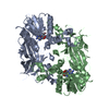
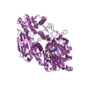
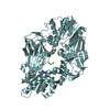
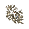
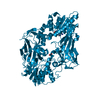
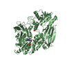
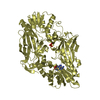
| Title | Crystal structure of Trypanosoma brucei protein arginine methyltransferase 7 complex with AdoHcy |
|---|
 Components Components | Protein arginine N-methyltransferase 7 |
|---|
 Keywords Keywords | TRANSFERASE / methyltransferase |
|---|
| Function / homology |  Function and homology information Function and homology information
arginine N-methyltransferase activity / protein-arginine omega-N monomethyltransferase activity / peptidyl-arginine N-methylation / peptidyl-arginine methylation / protein-arginine N-methyltransferase activity / histone methyltransferase activity / Transferases; Transferring one-carbon groups; Methyltransferases / chromatin remodeling / regulation of DNA-templated transcription / nucleolus ...arginine N-methyltransferase activity / protein-arginine omega-N monomethyltransferase activity / peptidyl-arginine N-methylation / peptidyl-arginine methylation / protein-arginine N-methyltransferase activity / histone methyltransferase activity / Transferases; Transferring one-carbon groups; Methyltransferases / chromatin remodeling / regulation of DNA-templated transcription / nucleolus / protein-containing complex / identical protein binding / cytoplasmSimilarity search - Function Ribosomal protein L11 methyltransferase (PrmA) / Hnrnp arginine n-methyltransferase1 / Hnrnp arginine n-methyltransferase1 / Protein arginine N-methyltransferase / SAM-dependent methyltransferase PRMT-type domain profile. / Vaccinia Virus protein VP39 / Distorted Sandwich / S-adenosyl-L-methionine-dependent methyltransferase superfamily / Rossmann fold / 3-Layer(aba) Sandwich ...Ribosomal protein L11 methyltransferase (PrmA) / Hnrnp arginine n-methyltransferase1 / Hnrnp arginine n-methyltransferase1 / Protein arginine N-methyltransferase / SAM-dependent methyltransferase PRMT-type domain profile. / Vaccinia Virus protein VP39 / Distorted Sandwich / S-adenosyl-L-methionine-dependent methyltransferase superfamily / Rossmann fold / 3-Layer(aba) Sandwich / Mainly Beta / Alpha BetaSimilarity search - Domain/homology |
|---|
| Biological species |   Trypanosoma brucei brucei (eukaryote) Trypanosoma brucei brucei (eukaryote) |
|---|
| Method |  X-RAY DIFFRACTION / MOLECULAR REPLACEMENT / Resolution: 1.7 Å X-RAY DIFFRACTION / MOLECULAR REPLACEMENT / Resolution: 1.7 Å |
|---|
 Authors Authors | Wang, C. / Zhu, Y. / Shi, Y. |
|---|
 Citation Citation | Journal: Structure / Year: 2014
Title: Structural determinants for the strict monomethylation activity by trypanosoma brucei protein arginine methyltransferase 7.
Authors: Wang, C. / Zhu, Y. / Caceres, T.B. / Liu, L. / Peng, J. / Wang, J. / Chen, J. / Chen, X. / Zhang, Z. / Zuo, X. / Gong, Q. / Teng, M. / Hevel, J.M. / Wu, J. / Shi, Y. |
|---|
| History | | Deposition | Aug 6, 2013 | Deposition site: RCSB / Processing site: PDBJ |
|---|
| Revision 1.0 | Apr 23, 2014 | Provider: repository / Type: Initial release |
|---|
| Revision 1.1 | Aug 24, 2022 | Group: Database references / Derived calculations / Category: citation / database_2 / struct_site
Item: _citation.journal_volume / _citation.page_first ..._citation.journal_volume / _citation.page_first / _citation.page_last / _citation.title / _database_2.pdbx_DOI / _database_2.pdbx_database_accession / _struct_site.pdbx_auth_asym_id / _struct_site.pdbx_auth_comp_id / _struct_site.pdbx_auth_seq_id |
|---|
| Revision 1.2 | May 29, 2024 | Group: Data collection / Category: chem_comp_atom / chem_comp_bond |
|---|
|
|---|
 Movie
Movie Controller

 Yorodumi
 Open data
 Basic information
 Structure visualization
 Downloads & links
 Other downloads


 PDBj
PDBj Assembly


 Type: L-peptide linking / Mass: 384.411 Da / Num. of mol.: 1 / Source method: obtained synthetically / Formula: C14H20N6O5S
 Mass: 18.015 Da / Num. of mol.: 246 / Source method: isolated from a natural source / Formula: H2O
 Sample preparation
 Processing