[English] 日本語
Yorodumi
- PDB-1nze: Crystal structure of PsbQ polypeptide of photosystem II from high... -

+
Open data


ID or keywords:

Loading...

-
Basic information

Entry
Database: PDB / ID: 1nze
Title
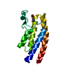
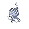
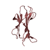
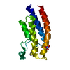
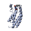
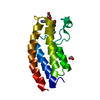
Crystal structure of PsbQ polypeptide of photosystem II from higher plants
ComponentsOxygen-evolving enhancer protein 3
KeywordsOXIDOREDUCTASE / Photosystem II / oxygen-enhancer evolving complex / water oxidizing complex / PsbQ / OEE3
Function / homology
Function and homology information


photosystem II oxygen evolving complex / photosynthetic electron transport chain / extrinsic component of membrane / electron transporter, transferring electrons within the cyclic electron transport pathway of photosynthesis activity / chloroplast thylakoid membrane / calcium ion binding
Similarity search - Function
Oxygen-evolving enhancer protein 3 (PsbQ), four-helix up-down bundle / Oxygen-evolving enhancer protein 3 / Oxygen evolving enhancer protein 3 / PsbQ-like domain superfamily / Four Helix Bundle (Hemerythrin (Met), subunit A) / Up-down Bundle / Mainly Alpha
Similarity search - Domain/homology
Oxygen-evolving enhancer protein 3, chloroplastic
Similarity search - Component
Biological speciesSpinacia oleracea (spinach)
MethodX-RAY DIFFRACTION / SYNCHROTRON / MAD / Resolution: 1.95 Å
AuthorsCalderone, V. / Trabucco, M. / Vujicic, A. / Battistutta, R. / Giacometti, G.M. / Andreucci, F. / Barbato, R. / Zanotti, G.
Citation
Journal: Embo Rep. / Year: 2003
Title: Crystal structure of the PsbQ protein of photosystem II from higher plants
Authors: Calderone, V. / Trabucco, M. / Vujicic, A. / Battistutta, R. / Giacometti, G.M. / Andreucci, F. / Barbato, R. / Zanotti, G.
#1: Journal: Proc.Natl.Acad.Sci.USA / Year: 2001
Title: Excited State Dynamics in Photosystem II: Insights from the X-Ray Crystal Structure
Authors: Vasilev, S. / Orth, P. / Zouni, A. / Owens, T.G. / Bruce, D.
#2: Journal: J.Biol.Chem. / Year: 2002
Title: Three-dimensional electron cryo-microscopy study of the extrinsic domains of the oxygen-evolving complex of spinach: assignment of the PsbO protein
Authors: Nield, J. / Balsera, M. / De Las Rivas, J. / Barber, J.
#3: Journal: Proc.Natl.Acad.Sci.USA / Year: 2003
Title: Crystal structure of oxygen-evolving photosystem II from Thermosynechoccus vulcanus at 3.7 A resolution
Authors: Kamiyam, N. / Shen, J.-R.
History
DepositionFeb 17, 2003Deposition site: RCSB / Processing site: RCSB
Revision 1.0Aug 26, 2003Provider: repository / Type: Initial release
Revision 1.1Apr 29, 2008Group: Version format compliance
Revision 1.2Jul 13, 2011Group: Version format compliance
Revision 1.3Feb 14, 2024Group: Data collection / Database references / Derived calculations
Category: chem_comp_atom / chem_comp_bond ...chem_comp_atom / chem_comp_bond / database_2 / pdbx_struct_conn_angle / struct_conn / struct_site
Item: _database_2.pdbx_DOI / _database_2.pdbx_database_accession ..._database_2.pdbx_DOI / _database_2.pdbx_database_accession / _pdbx_struct_conn_angle.ptnr1_auth_comp_id / _pdbx_struct_conn_angle.ptnr1_auth_seq_id / _pdbx_struct_conn_angle.ptnr1_label_asym_id / _pdbx_struct_conn_angle.ptnr1_label_atom_id / _pdbx_struct_conn_angle.ptnr1_label_comp_id / _pdbx_struct_conn_angle.ptnr1_label_seq_id / _pdbx_struct_conn_angle.ptnr1_symmetry / _pdbx_struct_conn_angle.ptnr3_auth_comp_id / _pdbx_struct_conn_angle.ptnr3_auth_seq_id / _pdbx_struct_conn_angle.ptnr3_label_asym_id / _pdbx_struct_conn_angle.ptnr3_label_atom_id / _pdbx_struct_conn_angle.ptnr3_label_comp_id / _pdbx_struct_conn_angle.ptnr3_label_seq_id / _pdbx_struct_conn_angle.ptnr3_symmetry / _pdbx_struct_conn_angle.value / _struct_conn.pdbx_dist_value / _struct_conn.ptnr1_auth_comp_id / _struct_conn.ptnr1_auth_seq_id / _struct_conn.ptnr1_label_asym_id / _struct_conn.ptnr1_label_atom_id / _struct_conn.ptnr1_label_comp_id / _struct_conn.ptnr1_label_seq_id / _struct_conn.ptnr1_symmetry / _struct_conn.ptnr2_auth_comp_id / _struct_conn.ptnr2_auth_seq_id / _struct_conn.ptnr2_label_asym_id / _struct_conn.ptnr2_label_atom_id / _struct_conn.ptnr2_label_comp_id / _struct_conn.ptnr2_label_seq_id / _struct_conn.ptnr2_symmetry / _struct_site.pdbx_auth_asym_id / _struct_site.pdbx_auth_comp_id / _struct_site.pdbx_auth_seq_id

-
Structure visualization

Structure viewerMolecule:
MolmilJmol/JSmol

Downloads & links

-
Assembly

Deposited unit
A: Oxygen-evolving enhancer protein 3
hetero molecules


Theoretical massNumber of molelcules
Total (without water)16,6102
Polymers16,5451
Non-polymers651
Water1,27971
1


  • Idetical with deposited unit
  • defined by author
TypeNameSymmetry operationNumber
identity operation1_555x,y,z1
Unit cell
Length a, b, c (Å)50.282, 50.282, 94.067
Angle α, β, γ (deg.)90.00, 90.00, 120.00
Int Tables number154
Space group name H-MP3221

-
Components

#1: Protein Oxygen-evolving enhancer protein 3 / OEE3 / 16 kDa subunit of oxygen evolving system of photosystem II / OEC 16 kDa subunit / PsbQ


Mass: 16544.746 Da / Num. of mol.: 1 / Source method: isolated from a natural source
Details: The protein was extracted from PS II thylacoid membranes
Source: (natural) Spinacia oleracea (spinach) / References: UniProt: P12301
#2: Chemical ChemComp-ZN / ZINC ION


Mass: 65.409 Da / Num. of mol.: 1 / Source method: obtained synthetically / Formula: Zn
#3: Water ChemComp-HOH / water


Mass: 18.015 Da / Num. of mol.: 71 / Source method: isolated from a natural source / Formula: H2O

-
Experimental details

-
Experiment

ExperimentMethod: X-RAY DIFFRACTION / Number of used crystals: 3

-
Sample preparation

CrystalDensity Matthews: 2.58 Å3/Da / Density % sol: 51.97 %
Crystal growTemperature: 298 K / Method: vapor diffusion, sitting drop / pH: 7.2
Details: 50 mM Tris, 50 mM NaCl, 5 mM Zn2+, pH 7.2, VAPOR DIFFUSION, SITTING DROP, temperature 298K
Crystal grow
*PLUS
Temperature: 20 ℃ / pH: 4.6 / Method: vapor diffusion
Components of the solutions
*PLUS
IDConc.Common nameCrystal-IDSol-IDDetailsChemical formula
120-24 mg/mlprotein1drop
225-30 %(w/v)PEG33501reservoiror PDB4000
35 mM1reservoirpH4.6Zn2+

-
Data collection

Diffraction
IDMean temperature (K)Crystal-ID
11001
21001
31001
Diffraction source
SourceSiteBeamlineIDWavelength (Å)
SYNCHROTRONESRF ID2910.93, 1.282, 1.284
SYNCHROTRONELETTRA 5.2R21.2
SYNCHROTRONESRF ID14-430.9393
Detector
TypeIDDetectorDate
ADSC QUANTUM 2101CCDFeb 15, 2002
MARRESEARCH2CCDSep 4, 2001
ADSC QUANTUM 43CCDApr 9, 2002
RadiationProtocol: MAD / Monochromatic (M) / Laue (L): M / Scattering type: x-ray
Radiation wavelength
IDWavelength (Å)Relative weight
10.931
21.2821
31.2841
41.21
50.93931
ReflectionResolution: 1.95→43.55 Å / Num. all: 10333 / Num. obs: 10333 / % possible obs: 98.1 % / Observed criterion σ(F): 0 / Observed criterion σ(I): 0 / Redundancy: 6.2 % / Biso Wilson estimate: 40.4 Å2 / Rmerge(I) obs: 0.11 / Rsym value: 0.11 / Net I/σ(I): 3.3
Reflection shellResolution: 1.95→2.06 Å / Redundancy: 5.3 % / Rmerge(I) obs: 0.33 / Mean I/σ(I) obs: 1.2 / Num. unique all: 1340 / Rsym value: 0.33 / % possible all: 90.9
Reflection
*PLUS
Lowest resolution: 39 Å / Redundancy: 8.9 %
Reflection shell
*PLUS
% possible obs: 90.9 % / Num. unique obs: 1340

-
Processing

Software
NameVersionClassification
CNS1refinement
MOSFLMdata reduction
CCP4(SCALA)data scaling
SOLVEphasing
RefinementMethod to determine structure: MAD / Resolution: 1.95→43.55 Å / Rfactor Rfree error: 0.008 / Isotropic thermal model: RESTRAINED / Cross valid method: THROUGHOUT / σ(F): 0 / σ(I): 0 / Stereochemistry target values: Engh & Huber
RfactorNum. reflection% reflectionSelection details
Rfree0.268 1062 10.3 %RANDOM
Rwork0.227 ---
all0.229 10333 --
obs0.227 10307 97.8 %-
Solvent computationSolvent model: FLAT MODEL / Bsol: 77.2503 Å2 / ksol: 0.368844 e/Å3
Displacement parametersBiso mean: 59.2 Å2
Baniso -1Baniso -2Baniso -3
1--4.56 Å22.22 Å20 Å2
2---6.75 Å20 Å2
3---11.31 Å2
Refine analyze
FreeObs
Luzzati coordinate error0.35 Å0.32 Å
Luzzati d res low-5 Å
Luzzati sigma a0.39 Å0.38 Å
Refinement stepCycle: LAST / Resolution: 1.95→43.55 Å
ProteinNucleic acidLigandSolventTotal
Num. atoms876 0 1 73 950
Refine LS restraints
Refine-IDTypeDev idealDev ideal target
X-RAY DIFFRACTIONc_bond_d0.009
X-RAY DIFFRACTIONc_bond_d_na
X-RAY DIFFRACTIONc_bond_d_prot
X-RAY DIFFRACTIONc_angle_d
X-RAY DIFFRACTIONc_angle_d_na
X-RAY DIFFRACTIONc_angle_d_prot
X-RAY DIFFRACTIONc_angle_deg1.5
X-RAY DIFFRACTIONc_angle_deg_na
X-RAY DIFFRACTIONc_angle_deg_prot
X-RAY DIFFRACTIONc_dihedral_angle_d20.3
X-RAY DIFFRACTIONc_dihedral_angle_d_na
X-RAY DIFFRACTIONc_dihedral_angle_d_prot
X-RAY DIFFRACTIONc_improper_angle_d0.97
X-RAY DIFFRACTIONc_improper_angle_d_na
X-RAY DIFFRACTIONc_improper_angle_d_prot
X-RAY DIFFRACTIONc_mcbond_it1.611.5
X-RAY DIFFRACTIONc_mcangle_it2.842
X-RAY DIFFRACTIONc_scbond_it2.422
X-RAY DIFFRACTIONc_scangle_it4.032.5
LS refinement shellResolution: 1.95→2.07 Å / Rfactor Rfree error: 0.031 / Total num. of bins used: 6
RfactorNum. reflection% reflection
Rfree0.387 158 10.3 %
Rwork0.382 1379 -
obs-1560 90 %
Xplor file
Refine-IDSerial noParam fileTopol file
X-RAY DIFFRACTION1PROTEIN_REP.PARAMPROTEIN.TOP
X-RAY DIFFRACTION2WATER_REP.PARAMWATER.TOP
X-RAY DIFFRACTION3ION.PARAMION.TOP
Refine LS restraints
*PLUS
Refine-IDTypeDev ideal
X-RAY DIFFRACTIONc_bond_d0.01
X-RAY DIFFRACTIONc_angle_deg1.5
X-RAY DIFFRACTIONc_dihedral_angle_d
X-RAY DIFFRACTIONc_dihedral_angle_deg20.3
X-RAY DIFFRACTIONc_improper_angle_d
X-RAY DIFFRACTIONc_improper_angle_deg0.97

+
About Yorodumi

-
News

-
Feb 9, 2022. New format data for meta-information of EMDB entries

New format data for meta-information of EMDB entries

  • Version 3 of the EMDB header file is now the official format.
  • The previous official version 1.9 will be removed from the archive.

Related info.:EMDB header

External links:wwPDB to switch to version 3 of the EMDB data model

-
Aug 12, 2020. Covid-19 info

Covid-19 info

URL: https://pdbjlvh1.pdbj.org/emnavi/covid19.php

New page: Covid-19 featured information page in EM Navigator.

Related info.:Covid-19 info / Mar 5, 2020. Novel coronavirus structure data

+
Mar 5, 2020. Novel coronavirus structure data

Novel coronavirus structure data

Related info.:Yorodumi Speices / Aug 12, 2020. Covid-19 info

External links:COVID-19 featured content - PDBj / Molecule of the Month (242):Coronavirus Proteases

+
Jan 31, 2019. EMDB accession codes are about to change! (news from PDBe EMDB page)

EMDB accession codes are about to change! (news from PDBe EMDB page)

  • The allocation of 4 digits for EMDB accession codes will soon come to an end. Whilst these codes will remain in use, new EMDB accession codes will include an additional digit and will expand incrementally as the available range of codes is exhausted. The current 4-digit format prefixed with “EMD-” (i.e. EMD-XXXX) will advance to a 5-digit format (i.e. EMD-XXXXX), and so on. It is currently estimated that the 4-digit codes will be depleted around Spring 2019, at which point the 5-digit format will come into force.
  • The EM Navigator/Yorodumi systems omit the EMD- prefix.

Related info.:Q: What is EMD? / ID/Accession-code notation in Yorodumi/EM Navigator

External links:EMDB Accession Codes are Changing Soon! / Contact to PDBj

+
Jul 12, 2017. Major update of PDB

Major update of PDB

  • wwPDB released updated PDB data conforming to the new PDBx/mmCIF dictionary.
  • This is a major update changing the version number from 4 to 5, and with Remediation, in which all the entries are updated.
  • In this update, many items about electron microscopy experimental information are reorganized (e.g. em_software).
  • Now, EM Navigator and Yorodumi are based on the updated data.

External links:wwPDB Remediation / Enriched Model Files Conforming to OneDep Data Standards Now Available in the PDB FTP Archive

-
Yorodumi

Thousand views of thousand structures

  • Yorodumi is a browser for structure data from EMDB, PDB, SASBDB, etc.
  • This page is also the successor to EM Navigator detail page, and also detail information page/front-end page for Omokage search.
  • The word "yorodu" (or yorozu) is an old Japanese word meaning "ten thousand". "mi" (miru) is to see.

Related info.:EMDB / PDB / SASBDB / Comparison of 3 databanks / Yorodumi Search / Aug 31, 2016. New EM Navigator & Yorodumi / Yorodumi Papers / Jmol/JSmol / Function and homology information / Changes in new EM Navigator and Yorodumi

Read more