 Movie
Movie Controller


+ Open data

- Basic information
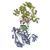
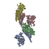
| Entry | Database: PDB / ID: 1fe1 | |||||||||
|---|---|---|---|---|---|---|---|---|---|---|
| Title | CRYSTAL STRUCTURE PHOTOSYSTEM II | |||||||||
 Components Components | (PROTEIN (PHOTOSYSTEM II: SUBUNIT ...) x 9 | |||||||||
 Keywords Keywords | PHOTOSYNTHESIS/ELECTRON TRANSPORT / PHOTOSYNTHESIS / PHOTOSYNTHETIC REACTION CENTER / CORE- ANTEN A LIGHT-HARVESTING SYSTEM / THERMOPHILIC CYANOBACTERIUM / MEMBRANE PROTEIN / PHOTOSYNTHESIS-ELECTRON TRANSPORT COMPLEX | |||||||||
| Function / homology | : / CHLOROPHYLL A / : / PROTOPORPHYRIN IX CONTAINING FE / : / PHEOPHYTIN A / Chem-PLA / TYROSINE Function and homology information Function and homology information | |||||||||
| Biological species |  Synechococcus elongatus (bacteria) Synechococcus elongatus (bacteria) | |||||||||
| Method |  X-RAY DIFFRACTION / SYNCHROTRON / MIRAS / Resolution: 3.8 Å X-RAY DIFFRACTION / SYNCHROTRON / MIRAS / Resolution: 3.8 Å | |||||||||
 Authors Authors | Zouni, A. / Witt, H.-T. / Kern, J. / Fromme, P. / Krauss, N. / Saenger, W. / Orth, P. | |||||||||
 Citation Citation | Journal: Nature / Year: 2001 Title: Crystal structure of photosystem II from Synechococcus elongatus at 3.8 A resolution. Authors: Zouni, A. / Witt, H.T. / Kern, J. / Fromme, P. / Krauss, N. / Saenger, W. / Orth, P. | |||||||||
| History |
|
- Structure visualization
| Structure viewer | Molecule: MolmilJmol/JSmol |
|---|
- Downloads & links
-Download
| PDBx/mmCIF format | 1fe1.cif.gz | 140.4 KB | Display | PDBx/mmCIF format |
|---|---|---|---|---|
| PDB format | pdb1fe1.ent.gz | 97.9 KB | Display | PDB format |
| PDBx/mmJSON format | 1fe1.json.gz | Tree view | PDBx/mmJSON format | |
| Others |  Other downloads Other downloads |
-Validation report
| Arichive directory | https://data.pdbj.org/pub/pdb/validation_reports/fe/1fe1ftp://data.pdbj.org/pub/pdb/validation_reports/fe/1fe1 | HTTPS FTP |
|---|
-Related structure data
| Similar structure data |
|---|


-Links
 PDBj
PDBj


- Assembly
| Deposited unit | 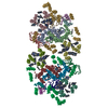
| ||||||||
|---|---|---|---|---|---|---|---|---|---|
| 1 |
| ||||||||
| Unit cell |
|
-Components
-PROTEIN (PHOTOSYSTEM II: SUBUNIT ... , 9 types, 18 molecules AJBKCLDMENFOGPHQIR
| #1: Protein | Mass: 14400.733 Da / Num. of mol.: 2 / Source method: isolated from a natural source / Source: (natural) Synechococcus elongatus (bacteria) / Cellular location: THYLAKOID MEMBRANE#2: Protein | Mass: 14826.253 Da / Num. of mol.: 2 / Source method: isolated from a natural source / Source: (natural) Synechococcus elongatus (bacteria) / Cellular location: THYLAKOID MEMBRANE#3: Protein | Mass: 13294.380 Da / Num. of mol.: 2 / Source method: isolated from a natural source / Source: (natural) Synechococcus elongatus (bacteria) / Cellular location: THYLAKOID MEMBRANE#4: Protein | Mass: 13209.274 Da / Num. of mol.: 2 / Source method: isolated from a natural source / Source: (natural) Synechococcus elongatus (bacteria) / Cellular location: THYLAKOID MEMBRANE#5: Protein/peptide | Mass: 3422.209 Da / Num. of mol.: 2 / Source method: isolated from a natural source / Source: (natural) Synechococcus elongatus (bacteria) / Cellular location: THYLAKOID MEMBRANE#6: Protein/peptide | Mass: 2571.161 Da / Num. of mol.: 2 / Source method: isolated from a natural source / Source: (natural) Synechococcus elongatus (bacteria) / Cellular location: THYLAKOID MEMBRANE#7: Protein | Mass: 26570.598 Da / Num. of mol.: 2 / Source method: isolated from a natural source / Source: (natural) Synechococcus elongatus (bacteria) / Cellular location: THYLAKOID MEMBRANE#8: Protein | Mass: 9805.078 Da / Num. of mol.: 2 / Source method: isolated from a natural source / Source: (natural) Synechococcus elongatus (bacteria) / Cellular location: THYLAKOID MEMBRANE, LUMEN#9: Protein | Mass: 7422.140 Da / Num. of mol.: 2 / Source method: isolated from a natural source / Source: (natural) Synechococcus elongatus (bacteria) / Cellular location: THYLAKOID MEMBRANE, LUMEN |
|---|
-Non-polymers , 8 types, 90 molecules 


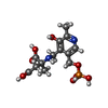


| #10: Chemical | ChemComp-MN /  Mass: 54.938 Da / Num. of mol.: 8 / Source method: obtained synthetically / Formula: Mn Mass: 54.938 Da / Num. of mol.: 8 / Source method: obtained synthetically / Formula: Mn#11: Chemical | ChemComp-TYR /  Type: L-peptide linking / Mass: 181.189 Da / Num. of mol.: 4 / Source method: obtained synthetically / Formula: C9H11NO3 Type: L-peptide linking / Mass: 181.189 Da / Num. of mol.: 4 / Source method: obtained synthetically / Formula: C9H11NO3#12: Chemical | ChemComp-CLA /  Mass: 893.489 Da / Num. of mol.: 64 / Source method: obtained synthetically / Formula: C55H72MgN4O5 Mass: 893.489 Da / Num. of mol.: 64 / Source method: obtained synthetically / Formula: C55H72MgN4O5#13: Chemical | ChemComp-PHO /  Mass: 871.200 Da / Num. of mol.: 4 / Source method: obtained synthetically / Formula: C55H74N4O5 Mass: 871.200 Da / Num. of mol.: 4 / Source method: obtained synthetically / Formula: C55H74N4O5#14: Chemical |  Mass: 55.845 Da / Num. of mol.: 2 / Source method: obtained synthetically / Formula: Fe Mass: 55.845 Da / Num. of mol.: 2 / Source method: obtained synthetically / Formula: Fe#15: Chemical |  Mass: 378.272 Da / Num. of mol.: 2 / Source method: obtained synthetically / Formula: C13H19N2O9P Mass: 378.272 Da / Num. of mol.: 2 / Source method: obtained synthetically / Formula: C13H19N2O9P#16: Chemical | ChemComp-HEM /  Mass: 616.487 Da / Num. of mol.: 4 / Source method: obtained synthetically / Formula: C34H32FeN4O4 Mass: 616.487 Da / Num. of mol.: 4 / Source method: obtained synthetically / Formula: C34H32FeN4O4#17: Chemical |  Mass: 112.411 Da / Num. of mol.: 2 / Source method: obtained synthetically / Formula: Cd Mass: 112.411 Da / Num. of mol.: 2 / Source method: obtained synthetically / Formula: Cd |
|---|
-Details
| Has protein modification | N |
|---|
-Experimental details
-Experiment
| Experiment | Method: X-RAY DIFFRACTION / Number of used crystals: 50 |
|---|
- Sample preparation
| Crystal | Density % sol: 45 % |
|---|---|
| Crystal grow | Temperature: 293 K / Method: batch method / pH: 7 Details: POLYETHYLENGLYCOL 2000, HEPES-BUFFER, CACL2 , pH 7.00, BATCH METHOD, temperature 293K |
| Crystal grow | *PLUS Method: unknown |
-Data collection
| Diffraction |
| |||||||||||||||||||||||||
|---|---|---|---|---|---|---|---|---|---|---|---|---|---|---|---|---|---|---|---|---|---|---|---|---|---|---|
| Diffraction source |
| |||||||||||||||||||||||||
| Detector |
| |||||||||||||||||||||||||
| Radiation | Protocol: SINGLE WAVELENGTH / Monochromatic (M) / Laue (L): M / Scattering type: x-ray | |||||||||||||||||||||||||
| Radiation wavelength | Wavelength: 0.9 Å / Relative weight: 1 | |||||||||||||||||||||||||
| Reflection | Resolution: 3.8→20 Å / Num. all: 84964 / Num. obs: 84964 / % possible obs: 95.4 % / Observed criterion σ(F): 0 / Observed criterion σ(I): 0 / Redundancy: 3.2 % / Biso Wilson estimate: 102 Å2 / Rmerge(I) obs: 0.068 / Net I/σ(I): 17.1 | |||||||||||||||||||||||||
| Reflection shell | Resolution: 3.8→3.91 Å / Redundancy: 2.5 % / Rmerge(I) obs: 0.418 / % possible all: 84.5 | |||||||||||||||||||||||||
| Reflection | *PLUS Highest resolution: 3.8 Å / Lowest resolution: 20 Å / Observed criterion σ(I): 0 / Redundancy: 3.2 % | |||||||||||||||||||||||||
| Reflection shell | *PLUS % possible obs: 84.5 % / Mean I/σ(I) obs: 3.4 |


- Processing
| Software |
| ||||||||||||
|---|---|---|---|---|---|---|---|---|---|---|---|---|---|
| Refinement | Method to determine structure: MIRAS / Resolution: 3.8→20 Å Details: THE STRUCTURE WAS SOLVED TO A RESOLUTION OF 4.2 ANGSTROEM USING 6 HEAVY ATOM DERIVATIVES. FOR DETAILS SEE ZOUNI ET AL. THE CADIMIUM DERIVATIVE HAS A RESOLUTION OF 3.8 ANGSTROEM. ...Details: THE STRUCTURE WAS SOLVED TO A RESOLUTION OF 4.2 ANGSTROEM USING 6 HEAVY ATOM DERIVATIVES. FOR DETAILS SEE ZOUNI ET AL. THE CADIMIUM DERIVATIVE HAS A RESOLUTION OF 3.8 ANGSTROEM. PHASEEXTENSION FROM 4.2 TILL 3.8 ANGSTROEM WAS PERFORMED USING TWO-FOLD NONCRYSTALLOGRAPHIC SYMMETRY AND SOLVENT FLATTENING. | ||||||||||||
| Refinement step | Cycle: LAST / Resolution: 3.8→20 Å
| ||||||||||||
| Refinement | *PLUS Rfactor obs: 0.59 | ||||||||||||
| Solvent computation | *PLUS | ||||||||||||
| Displacement parameters | *PLUS |
