 Movie
Movie Controller


+ Open data

- Basic information
| Entry | Database: PDB / ID: 1nyu | ||||||
|---|---|---|---|---|---|---|---|
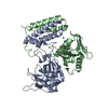
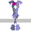
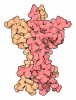
| Title | Crystal Structure of Activin A Bound to the ECD of ActRIIB | ||||||
 Components Components |
| ||||||
 Keywords Keywords | MEMBRANE PROTEIN/HORMONE/GROWTH FACTOR / ACTIVIN / TYPE II / TGF BETA / ACTRIIB / EXTRACELLULAR DOMAIN / MEMBRANE PROTEIN-HORMONE-GROWTH FACTOR COMPLEX | ||||||
| Function / homology |  Function and homology information Function and homology informationSignaling by BMP / progesterone secretion / activin A complex / inhibin A complex / cardiac fibroblast cell development / inhibin binding / negative regulation of B cell differentiation / GABAergic neuron differentiation / regulation of follicle-stimulating hormone secretion / Antagonism of Activin by Follistatin ...Signaling by BMP / progesterone secretion / activin A complex / inhibin A complex / cardiac fibroblast cell development / inhibin binding / negative regulation of B cell differentiation / GABAergic neuron differentiation / regulation of follicle-stimulating hormone secretion / Antagonism of Activin by Follistatin / TGFBR3 regulates activin signaling / negative regulation of follicle-stimulating hormone secretion / activin receptor activity / type II activin receptor binding / activin receptor activity, type II / lymphatic endothelial cell differentiation / striatal medium spiny neuron differentiation / positive regulation of ovulation / positive regulation of activin receptor signaling pathway / embryonic foregut morphogenesis / Glycoprotein hormones / positive regulation of follicle-stimulating hormone secretion / lymphangiogenesis / hemoglobin biosynthetic process / negative regulation of macrophage differentiation / gastrulation with mouth forming second / retina vasculature development in camera-type eye / Signaling by Activin / venous blood vessel development / cellular response to oxygen-glucose deprivation / positive regulation of extrinsic apoptotic signaling pathway in absence of ligand / eyelid development in camera-type eye / negative regulation of phosphorylation / organ growth / hair follicle development / artery development / cellular response to follicle-stimulating hormone stimulus / pattern specification process / cell surface receptor protein serine/threonine kinase signaling pathway / activin receptor complex / anterior/posterior pattern specification / SMAD protein signal transduction / receptor protein serine/threonine kinase / activin binding / cellular response to cholesterol / pancreas development / determination of left/right symmetry / transmembrane receptor protein serine/threonine kinase activity / Signaling by BMP / Signaling by Activin / activin receptor signaling pathway / negative regulation of ossification / negative regulation of activin receptor signaling pathway / skeletal system morphogenesis / odontogenesis of dentin-containing tooth / post-embryonic development / response to aldosterone / adrenal gland development / odontogenesis / insulin secretion / kinase activator activity / negative regulation of cold-induced thermogenesis / roof of mouth development / negative regulation of G1/S transition of mitotic cell cycle / endodermal cell differentiation / blood vessel remodeling / positive regulation of bone mineralization / growth factor binding / mesoderm development / ovarian follicle development / negative regulation of type II interferon production / positive regulation of transcription by RNA polymerase III / hematopoietic progenitor cell differentiation / positive regulation of osteoblast differentiation / lung development / positive regulation of collagen biosynthetic process / cellular response to angiotensin / positive regulation of protein metabolic process / peptide hormone binding / positive regulation of SMAD protein signal transduction / BMP signaling pathway / response to glucose / regulation of signal transduction / skeletal system development / extrinsic apoptotic signaling pathway / kidney development / positive regulation of erythrocyte differentiation / protein serine/threonine/tyrosine kinase activity / male gonad development / erythrocyte differentiation / response to activity / cytokine activity / growth factor activity / defense response / negative regulation of cell growth / hormone activity / cellular response to growth factor stimulus / cytokine-mediated signaling pathway / autophagy / heart development Similarity search - Function | ||||||
| Biological species |   Rattus norvegicus (Norway rat) Rattus norvegicus (Norway rat) Homo sapiens (human) Homo sapiens (human) | ||||||
| Method |  X-RAY DIFFRACTION / SYNCHROTRON / MOLECULAR REPLACEMENT / Resolution: 3.1 Å X-RAY DIFFRACTION / SYNCHROTRON / MOLECULAR REPLACEMENT / Resolution: 3.1 Å | ||||||
 Authors Authors | Thompson, T.B. / Woodruff, T.K. / Jardetzky, T.S. | ||||||
 Citation Citation | Journal: EMBO J. / Year: 2003 Title: Structures of an ActRIIB:activin A complex reveal a novel binding mode for TGF-beta ligand:receptor interactions Authors: Thompson, T.B. / Woodruff, T.K. / Jardetzky, T.S. | ||||||
| History |
|
- Structure visualization
| Structure viewer | Molecule: MolmilJmol/JSmol |
|---|
- Downloads & links
-Download
| PDBx/mmCIF format | 1nyu.cif.gz | 69.9 KB | Display | PDBx/mmCIF format |
|---|---|---|---|---|
| PDB format | pdb1nyu.ent.gz | 52 KB | Display | PDB format |
| PDBx/mmJSON format | 1nyu.json.gz | Tree view | PDBx/mmJSON format | |
| Others |  Other downloads Other downloads |
-Validation report
| Arichive directory | https://data.pdbj.org/pub/pdb/validation_reports/ny/1nyuftp://data.pdbj.org/pub/pdb/validation_reports/ny/1nyu | HTTPS FTP |
|---|
-Related structure data
| Related structure data |  1nysC 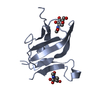 1bteS C: citing same article ( S: Starting model for refinement |
|---|---|
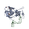
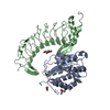
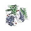
| Similar structure data |

-Links
 PDBj
PDBj
- Assembly
| Deposited unit | 
| ||||||||
|---|---|---|---|---|---|---|---|---|---|
| 1 |
| ||||||||
| Unit cell |
|
-Components
| #1: Protein | Mass: 12341.596 Da / Num. of mol.: 2 / Fragment: N-terminal Extracellular Domain (residues 19-119) Source method: isolated from a genetically manipulated source Source: (gene. exp.) Rattus norvegicus (Norway rat) / Gene: actrIIb / Plasmid: pvl1392 / Production host:   Spodoptera frugiperda (fall armyworm) / Strain (production host): SF+ / References: UniProt: P38445 Spodoptera frugiperda (fall armyworm) / Strain (production host): SF+ / References: UniProt: P38445#2: Protein | Mass: 12991.865 Da / Num. of mol.: 2 / Fragment: Mature Domain (residues 311-426) Source method: isolated from a genetically manipulated source Source: (gene. exp.) Homo sapiens (human) / Gene: INHBA / Production host:  Cricetulus griseus (Chinese hamster) / Strain (production host): BA83.6-02 / References: UniProt: P08476 Cricetulus griseus (Chinese hamster) / Strain (production host): BA83.6-02 / References: UniProt: P08476Has protein modification | Y | |
|---|
-Experimental details
-Experiment
| Experiment | Method: X-RAY DIFFRACTION / Number of used crystals: 1 |
|---|
- Sample preparation
| Crystal | Density Matthews: 3.45 Å3/Da / Density % sol: 64.11 % | |||||||||||||||||||||||||||||||||||||||||||||||||
|---|---|---|---|---|---|---|---|---|---|---|---|---|---|---|---|---|---|---|---|---|---|---|---|---|---|---|---|---|---|---|---|---|---|---|---|---|---|---|---|---|---|---|---|---|---|---|---|---|---|---|
| Crystal grow | Temperature: 295 K / Method: vapor diffusion, hanging drop / pH: 7 Details: PEG 4000, sodium chloride, hepes, pH 7.0, VAPOR DIFFUSION, HANGING DROP, temperature 295K | |||||||||||||||||||||||||||||||||||||||||||||||||
| Crystal grow | *PLUS pH: 7.5 | |||||||||||||||||||||||||||||||||||||||||||||||||
| Components of the solutions | *PLUS
|
-Data collection
| Diffraction | Mean temperature: 113 K |
|---|---|
| Diffraction source | Source: SYNCHROTRON / Site: APS  / Beamline: 5ID-B / Wavelength: 1 Å / Beamline: 5ID-B / Wavelength: 1 Å |
| Detector | Type: MARRESEARCH / Detector: CCD / Date: Apr 28, 2002 |
| Radiation | Protocol: SINGLE WAVELENGTH / Monochromatic (M) / Laue (L): M / Scattering type: x-ray |
| Radiation wavelength | Wavelength: 1 Å / Relative weight: 1 |
| Reflection | Resolution: 3.1→14.99 Å / Num. obs: 8777 / % possible obs: 99.5 % / Redundancy: 14.5 % / Biso Wilson estimate: 95.8 Å2 / Rmerge(I) obs: 0.083 / Net I/σ(I): 31.1 |
| Reflection shell | Resolution: 3.1→3.15 Å / Rmerge(I) obs: 0.437 / Mean I/σ(I) obs: 5.9 |
| Reflection | *PLUS Lowest resolution: 15 Å / Num. obs: 8815 / % possible obs: 97.6 % / Num. measured all: 128261 |
| Reflection shell | *PLUS % possible obs: 95.6 % |
- Processing
| Software |
| |||||||||||||||||||||||||
|---|---|---|---|---|---|---|---|---|---|---|---|---|---|---|---|---|---|---|---|---|---|---|---|---|---|---|
| Refinement | Method to determine structure: MOLECULAR REPLACEMENT Starting model: PDB ENTRY 1BTE Resolution: 3.1→14.99 Å / Rfactor Rfree error: 0.011 / Isotropic thermal model: RESTRAINED / Cross valid method: THROUGHOUT / σ(F): 0
| |||||||||||||||||||||||||
| Solvent computation | Solvent model: FLAT MODEL / Bsol: 34.2202 Å2 / ksol: 0.262393 e/Å3 | |||||||||||||||||||||||||
| Displacement parameters | Biso mean: 95.8 Å2
| |||||||||||||||||||||||||
| Refine analyze |
| |||||||||||||||||||||||||
| Refinement step | Cycle: LAST / Resolution: 3.1→14.99 Å
| |||||||||||||||||||||||||
| Refine LS restraints |
| |||||||||||||||||||||||||
| LS refinement shell | Resolution: 3.1→3.29 Å / Rfactor Rfree error: 0.034 / Total num. of bins used: 6
| |||||||||||||||||||||||||
| Xplor file |
| |||||||||||||||||||||||||
| Refinement | *PLUS % reflection Rfree: 5 % | |||||||||||||||||||||||||
| Solvent computation | *PLUS | |||||||||||||||||||||||||
| Displacement parameters | *PLUS | |||||||||||||||||||||||||
| Refine LS restraints | *PLUS
| |||||||||||||||||||||||||
| LS refinement shell | *PLUS Lowest resolution: 3.15 Å |
