 Movie
Movie Controller

[English] 日本語
 Yorodumi
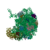
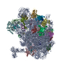
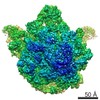
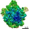
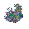
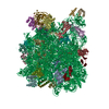
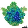
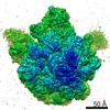
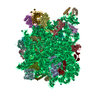
- PDB-1nwy: COMPLEX OF THE LARGE RIBOSOMAL SUBUNIT FROM DEINOCOCCUS RADIODURA... -
+ Open data

- Basic information
| Entry | Database: PDB / ID: 1nwy | ||||||
|---|---|---|---|---|---|---|---|
| Title | COMPLEX OF THE LARGE RIBOSOMAL SUBUNIT FROM DEINOCOCCUS RADIODURANS WITH AZITHROMYCIN | ||||||
 Components Components |
| ||||||
 Keywords Keywords | RIBOSOME / AZITHROMYCIN / MACROLIDE / KETOLIDE / 50S / RIBOSOMAL | ||||||
| Function / homology |  Function and homology information Function and homology informationlarge ribosomal subunit / transferase activity / 5S rRNA binding / ribosomal large subunit assembly / large ribosomal subunit rRNA binding / cytosolic large ribosomal subunit / cytoplasmic translation / tRNA binding / negative regulation of translation / rRNA binding ...large ribosomal subunit / transferase activity / 5S rRNA binding / ribosomal large subunit assembly / large ribosomal subunit rRNA binding / cytosolic large ribosomal subunit / cytoplasmic translation / tRNA binding / negative regulation of translation / rRNA binding / structural constituent of ribosome / ribosome / translation / ribonucleoprotein complex / mRNA binding / RNA binding / zinc ion binding / metal ion binding / cytoplasm Similarity search - Function | ||||||
| Biological species |  Deinococcus radiodurans (radioresistant) Deinococcus radiodurans (radioresistant) | ||||||
| Method |  X-RAY DIFFRACTION / SYNCHROTRON / FOURIER SYNTHESIS / Resolution: 3.3 Å X-RAY DIFFRACTION / SYNCHROTRON / FOURIER SYNTHESIS / Resolution: 3.3 Å | ||||||
 Authors Authors | Schluenzen, F. / Harms, J. / Franceschi, F. / Hansen, H.A.S. / Bartels, H. / Zarivach, R. / Yonath, A. | ||||||
 Citation Citation | Journal: Structure / Year: 2003 Title: Structural basis for the antibiotic activity of ketolides and azalides. Authors: Schlunzen, F. / Harms, J.M. / Franceschi, F. / Hansen, H.A. / Bartels, H. / Zarivach, R. / Yonath, A. | ||||||
| History |
|
- Structure visualization
| Structure viewer | Molecule: MolmilJmol/JSmol |
|---|
- Downloads & links
-Download
| PDBx/mmCIF format | 1nwy.cif.gz | 1.5 MB | Display | PDBx/mmCIF format |
|---|---|---|---|---|
| PDB format | pdb1nwy.ent.gz | 1.1 MB | Display | PDB format |
| PDBx/mmJSON format | 1nwy.json.gz | Tree view | PDBx/mmJSON format | |
| Others |  Other downloads Other downloads |
-Validation report
| Arichive directory | https://data.pdbj.org/pub/pdb/validation_reports/nw/1nwyftp://data.pdbj.org/pub/pdb/validation_reports/nw/1nwy | HTTPS FTP |
|---|
-Related structure data
| Related structure data |  1nwxC  1lnr S: Starting model for refinement C: citing same article ( |
|---|---|
| Similar structure data |

-Links
 PDBj
PDBj


- Assembly
| Deposited unit | 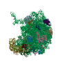
| ||||||||
|---|---|---|---|---|---|---|---|---|---|
| 1 |
| ||||||||
| Unit cell |
|
-Components
-RNA chain , 2 types, 2 molecules 09
| #1: RNA chain | Mass: 933405.000 Da / Num. of mol.: 1 / Source method: isolated from a natural source / Source: (natural) Deinococcus radiodurans (radioresistant) / References: GenBank: 15805042 |
|---|---|
| #2: RNA chain | Mass: 39911.859 Da / Num. of mol.: 1 / Source method: isolated from a natural source / Source: (natural) Deinococcus radiodurans (radioresistant) / References: GenBank: 15805042 |
+Ribosomal protein ... , 28 types, 28 molecules ABCDEFGHIJKLMNOPQRSUWXYZ1234
-Protein / Non-polymers , 2 types, 3 molecules T
| #22: Protein | Mass: 25415.418 Da / Num. of mol.: 1 / Source method: isolated from a natural source / Source: (natural) Deinococcus radiodurans (radioresistant) / References: UniProt: Q9RX88 |
|---|---|
| #32: Chemical |  Mass: 748.984 Da / Num. of mol.: 2 / Source method: obtained synthetically / Formula: C38H72N2O12 / Comment: antibiotic*YM Mass: 748.984 Da / Num. of mol.: 2 / Source method: obtained synthetically / Formula: C38H72N2O12 / Comment: antibiotic*YM |
-Experimental details
-Experiment
| Experiment | Method: X-RAY DIFFRACTION / Number of used crystals: 1 |
|---|
- Sample preparation
| Crystal | Density Matthews: 4 Å3/Da / Density % sol: 64 % | |||||||||||||||||||||||||||||||||||
|---|---|---|---|---|---|---|---|---|---|---|---|---|---|---|---|---|---|---|---|---|---|---|---|---|---|---|---|---|---|---|---|---|---|---|---|---|
| Crystal grow | Temperature: 291 K / Method: vapor diffusion, hanging drop / pH: 7.8 Details: ETHANOL, DIMETHYLHEXANEDIOL, MGCL2, KCL, HEPES, NH4CL, pH 7.80, VAPOR DIFFUSION, HANGING DROP, temperature 291K | |||||||||||||||||||||||||||||||||||
| Crystal grow | *PLUS Temperature: 18 ℃ / pH: 7.8 / Method: vapor diffusionDetails: Harms, J.M., (2001) Cell (Cambridge,Mass.), 107, 679. | |||||||||||||||||||||||||||||||||||
| Components of the solutions | *PLUS
|
-Data collection
| Diffraction | Mean temperature: 90 K |
|---|---|
| Diffraction source | Source: SYNCHROTRON / Site: APS  / Beamline: 19-ID / Wavelength: 1.0332 / Wavelength: 1.0332 Å / Beamline: 19-ID / Wavelength: 1.0332 / Wavelength: 1.0332 Å |
| Detector | Type: SBC / Detector: CCD / Date: Nov 14, 2001 |
| Radiation | Monochromator: SI111 OR SI311 / Protocol: SINGLE WAVELENGTH / Monochromatic (M) / Laue (L): M / Scattering type: x-ray |
| Radiation wavelength | Wavelength: 1.0332 Å / Relative weight: 1 |
| Reflection | Resolution: 3.2→30 Å / Num. all: 330873 / Num. obs: 330873 / % possible obs: 86.6 % / Observed criterion σ(F): 0 / Observed criterion σ(I): 0 / Redundancy: 4.2 % / Rsym value: 0.11 / Net I/σ(I): 9 |
| Reflection shell | Resolution: 3.2→3.25 Å / Redundancy: 1.7 % / Mean I/σ(I) obs: 1.9 / Rsym value: 0.34 / % possible all: 83.4 |
| Reflection | *PLUS Highest resolution: 3.2 Å / Lowest resolution: 30 Å / Rmerge(I) obs: 0.11 |
| Reflection shell | *PLUS % possible obs: 83.4 % / Rmerge(I) obs: 0.339 |
- Processing
| Software |
| ||||||||||||||||||||||||||||||||||||||||||||||||||||||||||||
|---|---|---|---|---|---|---|---|---|---|---|---|---|---|---|---|---|---|---|---|---|---|---|---|---|---|---|---|---|---|---|---|---|---|---|---|---|---|---|---|---|---|---|---|---|---|---|---|---|---|---|---|---|---|---|---|---|---|---|---|---|---|
| Refinement | Method to determine structure: FOURIER SYNTHESIS Starting model: PDB ENTRY 1LNR 1lnr Resolution: 3.3→15 Å / σ(F): 0 / Stereochemistry target values: ENGH & HUBER Details: THE STRUCTURE CONTAINS CA COORDINATES ONLY FOR THE PROTEIN CHAINS.
| ||||||||||||||||||||||||||||||||||||||||||||||||||||||||||||
| Refinement step | Cycle: LAST / Resolution: 3.3→15 Å
| ||||||||||||||||||||||||||||||||||||||||||||||||||||||||||||
| Refine LS restraints |
| ||||||||||||||||||||||||||||||||||||||||||||||||||||||||||||
| Refinement | *PLUS Highest resolution: 3.2 Å / Lowest resolution: 30 Å | ||||||||||||||||||||||||||||||||||||||||||||||||||||||||||||
| Solvent computation | *PLUS | ||||||||||||||||||||||||||||||||||||||||||||||||||||||||||||
| Displacement parameters | *PLUS |
