 Movie
Movie Controller

[English] 日本語
 Yorodumi
- PDB-1npc: THE STRUCTURE OF NEUTRAL PROTEASE FROM BACILLUS CEREUS AT 0.2-NM ... -
+ Open data

- Basic information
| Entry | Database: PDB / ID: 1npc | ||||||
|---|---|---|---|---|---|---|---|
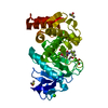
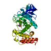
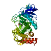
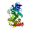
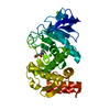
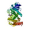
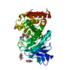
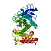
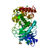
| Title | THE STRUCTURE OF NEUTRAL PROTEASE FROM BACILLUS CEREUS AT 0.2-NM RESOLUTION | ||||||
 Components Components | NEUTRAL PROTEASE | ||||||
 Keywords Keywords | HYDROLASE(METALLOPROTEINASE) | ||||||
| Function / homology |  Function and homology information Function and homology informationbacillolysin / metalloendopeptidase activity / proteolysis / extracellular region / metal ion binding Similarity search - Function | ||||||
| Biological species |  | ||||||
| Method |  X-RAY DIFFRACTION / Resolution: 2 Å X-RAY DIFFRACTION / Resolution: 2 Å | ||||||
 Authors Authors | Stark, W. / Pauptit, R.A. / Jansonius, J.N. | ||||||
 Citation Citation | Journal: Eur.J.Biochem. / Year: 1992 Title: The structure of neutral protease from Bacillus cereus at 0.2-nm resolution. Authors: Stark, W. / Pauptit, R.A. / Wilson, K.S. / Jansonius, J.N. #1: Journal: J.Mol.Biol. / Year: 1988Title: Crystal Structure of Neutral Protease from Bacillus Cereus Refined at 3.0 Angstroms Resolution and Comparison with the Homologous But More Thermostable Enzyme Thermolysin Authors: Pauptit, R.A. / Karlsson, R. / Picot, D. / Jenkins, J.A. / Niklaus-Reimer, A. / Jansonius, J.N. #2: Journal: Biol.Chem.Hoppe-Seyler / Year: 1986Title: The Primary Structure of Bacillus Cereus Neutral Proteinase and Comparison with Thermolysin and Bacillus Subtilis Proteinase Authors: Sidler, W. / Niederer, E. / Suter, F. / Zuber, H. | ||||||
| History |
|
- Structure visualization
| Structure viewer | Molecule: MolmilJmol/JSmol |
|---|
- Downloads & links
-Download
| PDBx/mmCIF format | 1npc.cif.gz | 80.8 KB | Display | PDBx/mmCIF format |
|---|---|---|---|---|
| PDB format | pdb1npc.ent.gz | 59.5 KB | Display | PDB format |
| PDBx/mmJSON format | 1npc.json.gz | Tree view | PDBx/mmJSON format | |
| Others |  Other downloads Other downloads |
-Validation report
| Arichive directory | https://data.pdbj.org/pub/pdb/validation_reports/np/1npcftp://data.pdbj.org/pub/pdb/validation_reports/np/1npc | HTTPS FTP |
|---|
-Related structure data
| Similar structure data |
|---|


-Links
 PDBj
PDBj- Assembly
| Deposited unit | 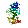
| |||||||||||||||
|---|---|---|---|---|---|---|---|---|---|---|---|---|---|---|---|---|
| 1 |
| |||||||||||||||
| Unit cell |
| |||||||||||||||
| Atom site foot note | 1: RESIDUE 52 IS A CIS PROLINE. | |||||||||||||||
| Components on special symmetry positions |
|
-Components
| #1: Protein | Mass: 33816.504 Da / Num. of mol.: 1 Source method: isolated from a genetically manipulated source Source: (gene. exp.) | ||||
|---|---|---|---|---|---|
| #2: Chemical | ChemComp-CA /   Mass: 40.078 Da / Num. of mol.: 4 / Source method: obtained synthetically / Formula: Ca Mass: 40.078 Da / Num. of mol.: 4 / Source method: obtained synthetically / Formula: Ca#3: Chemical | ChemComp-ZN / |   Mass: 65.409 Da / Num. of mol.: 1 / Source method: obtained synthetically / Formula: Zn Mass: 65.409 Da / Num. of mol.: 1 / Source method: obtained synthetically / Formula: Zn#4: Water | ChemComp-HOH / |  Mass: 18.015 Da / Num. of mol.: 304 / Source method: isolated from a natural source / Formula: H2O Mass: 18.015 Da / Num. of mol.: 304 / Source method: isolated from a natural source / Formula: H2O |
-Experimental details
-Experiment
| Experiment | Method: X-RAY DIFFRACTION |
|---|
- Sample preparation
| Crystal | Density Matthews: 2.51 Å3/Da / Density % sol: 50.99 % | |||||||||||||||||||||||||
|---|---|---|---|---|---|---|---|---|---|---|---|---|---|---|---|---|---|---|---|---|---|---|---|---|---|---|
| Crystal grow | *PLUS Temperature: 0 K / pH: 6 / Method: vapor diffusion, hanging drop | |||||||||||||||||||||||||
| Components of the solutions | *PLUS
|
-Data collection
| Radiation | Scattering type: x-ray |
|---|---|
| Radiation wavelength | Relative weight: 1 |
| Reflection | *PLUS Num. obs: 21168 / % possible obs: 88 % / Num. measured all: 195223 / Rmerge(I) obs: 0.219 |
- Processing
| Software | Name: TNT / Classification: refinement | ||||||||||||||||||||||||||||||
|---|---|---|---|---|---|---|---|---|---|---|---|---|---|---|---|---|---|---|---|---|---|---|---|---|---|---|---|---|---|---|---|
| Refinement | Resolution: 2→10 Å /
| ||||||||||||||||||||||||||||||
| Refinement step | Cycle: LAST / Resolution: 2→10 Å
| ||||||||||||||||||||||||||||||
| Refine LS restraints |
| ||||||||||||||||||||||||||||||
| Refinement | *PLUS Highest resolution: 2 Å / Lowest resolution: 10 Å / Num. reflection obs: 21168 / Rfactor obs: 0.175 | ||||||||||||||||||||||||||||||
| Solvent computation | *PLUS | ||||||||||||||||||||||||||||||
| Displacement parameters | *PLUS | ||||||||||||||||||||||||||||||
| Refine LS restraints | *PLUS
|
