 Movie
Movie Controller

[English] 日本語
 Yorodumi
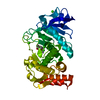
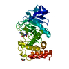
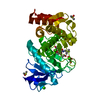
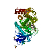
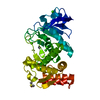
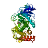
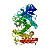
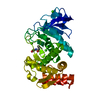
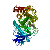
- PDB-1z9g: Crystal Structure Analysis of Thermolysin Complexed with the Inhi... -
+ Open data

- Basic information
| Entry | Database: PDB / ID: 1z9g | ||||||
|---|---|---|---|---|---|---|---|
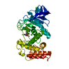
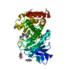
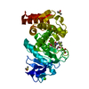
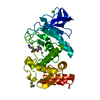
| Title | Crystal Structure Analysis of Thermolysin Complexed with the Inhibitor (R)-retro-thiorphan | ||||||
 Components Components | Thermolysin | ||||||
 Keywords Keywords | HYDROLASE / enzyme-inhibitor complex / zinc endopeptidase / gamma turn / thermostable | ||||||
| Function / homology |  Function and homology information Function and homology informationthermolysin / metalloendopeptidase activity / proteolysis / extracellular region / metal ion binding Similarity search - Function | ||||||
| Biological species |  | ||||||
| Method |  X-RAY DIFFRACTION / FOURIER SYNTHESIS / Resolution: 1.7 Å X-RAY DIFFRACTION / FOURIER SYNTHESIS / Resolution: 1.7 Å | ||||||
 Authors Authors | Roderick, S.L. / Fournie-Zaluski, M.C. / Roques, B.P. / Matthews, B.W. | ||||||
 Citation Citation | Journal: Biochemistry / Year: 1989 Title: Thiorphan and retro-thiorphan display equivalent interactions when bound to crystalline thermolysin Authors: Roderick, S.L. / Fournie-Zaluski, M.C. / Roques, B.P. / Matthews, B.W. | ||||||
| History |
|
- Structure visualization
| Structure viewer | Molecule: MolmilJmol/JSmol |
|---|
- Downloads & links
-Download
| PDBx/mmCIF format | 1z9g.cif.gz | 81.8 KB | Display | PDBx/mmCIF format |
|---|---|---|---|---|
| PDB format | pdb1z9g.ent.gz | 59.1 KB | Display | PDB format |
| PDBx/mmJSON format | 1z9g.json.gz | Tree view | PDBx/mmJSON format | |
| Others |  Other downloads Other downloads |
-Validation report
| Arichive directory | https://data.pdbj.org/pub/pdb/validation_reports/z9/1z9gftp://data.pdbj.org/pub/pdb/validation_reports/z9/1z9g | HTTPS FTP |
|---|
-Related structure data
| Related structure data |  1zdpC  3tln S: Starting model for refinement C: citing same article ( |
|---|---|
| Similar structure data |


-Links
 PDBj
PDBj- Assembly
| Deposited unit | 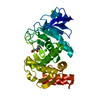
| ||||||||||
|---|---|---|---|---|---|---|---|---|---|---|---|
| 1 |
| ||||||||||
| Unit cell |
| ||||||||||
| Details | The enzyme acts as a monomer. |
-Components
| #1: Protein | Mass: 34362.305 Da / Num. of mol.: 1 / Source method: isolated from a natural source / Source: (natural) | ||||||
|---|---|---|---|---|---|---|---|
| #2: Chemical | ChemComp-CA /   Mass: 40.078 Da / Num. of mol.: 4 / Source method: obtained synthetically / Formula: Ca Mass: 40.078 Da / Num. of mol.: 4 / Source method: obtained synthetically / Formula: Ca#3: Chemical | ChemComp-ZN / |   Mass: 65.409 Da / Num. of mol.: 1 / Source method: obtained synthetically / Formula: Zn Mass: 65.409 Da / Num. of mol.: 1 / Source method: obtained synthetically / Formula: Zn#4: Chemical | ChemComp-RRT / ( |   Mass: 253.317 Da / Num. of mol.: 1 / Source method: obtained synthetically / Formula: C12H15NO3S Mass: 253.317 Da / Num. of mol.: 1 / Source method: obtained synthetically / Formula: C12H15NO3S#5: Water | ChemComp-HOH / |  Mass: 18.015 Da / Num. of mol.: 161 / Source method: isolated from a natural source / Formula: H2O Mass: 18.015 Da / Num. of mol.: 161 / Source method: isolated from a natural source / Formula: H2O |
-Experimental details
-Experiment
| Experiment | Method: X-RAY DIFFRACTION / Number of used crystals: 1 |
|---|
- Sample preparation
| Crystal | Density Matthews: 2.43 Å3/Da / Density % sol: 49 % |
|---|---|
| Crystal grow | Temperature: 277 K / Method: dilution with h2o / pH: 7.2 Details: Tris acetate, calcium acetate, DMSO, pH 7.2, dilution with H2O, temperature 277K |
-Data collection
| Diffraction | Mean temperature: 298 K |
|---|---|
| Diffraction source | Source: ROTATING ANODE / Type: ELLIOTT GX-21 / Wavelength: 1.5418 Å |
| Detector | Type: KODAK / Detector: FILM / Date: 1987 / Details: graphite monochromator, collimating slits |
| Radiation | Monochromator: graphite / Protocol: SINGLE WAVELENGTH / Monochromatic (M) / Laue (L): M / Scattering type: x-ray |
| Radiation wavelength | Wavelength: 1.5418 Å / Relative weight: 1 |
| Reflection | Resolution: 1.7→30 Å / Num. all: 27574 / Num. obs: 27574 / % possible obs: 72 % / Observed criterion σ(F): 0 / Observed criterion σ(I): 0 / Redundancy: 2.8 % / Biso Wilson estimate: 15.5 Å2 / Rmerge(I) obs: 0.058 / Rsym value: 0.032 |
- Processing
| Software |
| ||||||||||||||||||||||||||||||||||||
|---|---|---|---|---|---|---|---|---|---|---|---|---|---|---|---|---|---|---|---|---|---|---|---|---|---|---|---|---|---|---|---|---|---|---|---|---|---|
| Refinement | Method to determine structure: FOURIER SYNTHESIS Starting model: PDB ENTRY 3TLN 3tln Resolution: 1.7→10 Å / Isotropic thermal model: isotropic / Cross valid method: NONE / σ(F): 0 / σ(I): 0 / Stereochemistry target values: TNT version 1.0 / Details: 40 cycles of conjugate gradient refinement
| ||||||||||||||||||||||||||||||||||||
| Refinement step | Cycle: LAST / Resolution: 1.7→10 Å
| ||||||||||||||||||||||||||||||||||||
| Refine LS restraints |
|
