 Movie
Movie Controller


+ Open data

- Basic information
| Entry | Database: PDB / ID: 1noz | ||||||
|---|---|---|---|---|---|---|---|
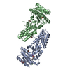
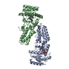
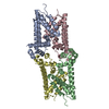
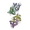
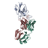
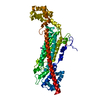
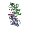
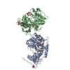
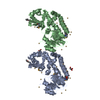
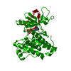
| Title | T4 DNA POLYMERASE FRAGMENT (RESIDUES 1-388) AT 110K | ||||||
 Components Components | DNA POLYMERASE | ||||||
 Keywords Keywords | NUCLEOTIDYLTRANSFERASE / EXONUCLEASE / DNA-BINDING | ||||||
| Function / homology |  Function and homology information Function and homology informationbidirectional double-stranded viral DNA replication / Hydrolases; Acting on ester bonds; Exodeoxyribonucleases producing 5'-phosphomonoesters / 3'-5' exonuclease activity / DNA-templated DNA replication / DNA-directed DNA polymerase / DNA-directed DNA polymerase activity / nucleotide binding / DNA binding / metal ion binding Similarity search - Function | ||||||
| Biological species |  Enterobacteria phage T4 (virus) Enterobacteria phage T4 (virus) | ||||||
| Method |  X-RAY DIFFRACTION / Resolution: 2.2 Å X-RAY DIFFRACTION / Resolution: 2.2 Å | ||||||
 Authors Authors | Wang, J. / Yu, P. / Lin, T.C. / Konigsberg, W.H. / Steitz, T.A. | ||||||
 Citation Citation | Journal: Biochemistry / Year: 1996 Title: Crystal structures of an NH2-terminal fragment of T4 DNA polymerase and its complexes with single-stranded DNA and with divalent metal ions. Authors: Wang, J. / Yu, P. / Lin, T.C. / Konigsberg, W.H. / Steitz, T.A. #1: Journal: J.Biol.Chem. / Year: 1994Title: Isolation, Characterization, and Kinetic Properties of Truncated Forms of T4 DNA Polymerase that Exhibit 3'-5' Exonuclease Activity Authors: Lin, T.C. / Karam, G. / Konigsberg, W.H. #2: Journal: J.Biol.Chem. / Year: 1988Title: Primary Structure of T4 DNA Polymerase. Evolutionary Relatedness to Eucaryotic and Other Procaryotic DNA Polymerases Authors: Spicer, E.K. / Rush, J. / Fung, C. / Reha-Krantz, L.J. / Karam, J.D. / Konigsberg, W.H. | ||||||
| History |
|
- Structure visualization
| Structure viewer | Molecule: MolmilJmol/JSmol |
|---|
- Downloads & links
-Download
| PDBx/mmCIF format | 1noz.cif.gz | 150.2 KB | Display | PDBx/mmCIF format |
|---|---|---|---|---|
| PDB format | pdb1noz.ent.gz | 119.8 KB | Display | PDB format |
| PDBx/mmJSON format | 1noz.json.gz | Tree view | PDBx/mmJSON format | |
| Others |  Other downloads Other downloads |
-Validation report
| Arichive directory | https://data.pdbj.org/pub/pdb/validation_reports/no/1nozftp://data.pdbj.org/pub/pdb/validation_reports/no/1noz | HTTPS FTP |
|---|
-Related structure data

-Links
 PDBj
PDBj


- Assembly
| Deposited unit | 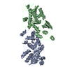
| ||||||||
|---|---|---|---|---|---|---|---|---|---|
| 1 |
| ||||||||
| 2 | 
| ||||||||
| Unit cell |
|
-Components
| #1: Protein | Mass: 45315.727 Da / Num. of mol.: 2 / Fragment: RESIDUES 1 - 388 Source method: isolated from a genetically manipulated source Source: (gene. exp.) Enterobacteria phage T4 (virus) / Genus: T4-like viruses / Species: Enterobacteria phage T4 sensu lato / Production host:  #2: Water | ChemComp-HOH / |  Mass: 18.015 Da / Num. of mol.: 45 / Source method: isolated from a natural source / Formula: H2O Mass: 18.015 Da / Num. of mol.: 45 / Source method: isolated from a natural source / Formula: H2OHas protein modification | Y | |
|---|
-Experimental details
-Experiment
| Experiment | Method: X-RAY DIFFRACTION |
|---|
- Sample preparation
| Crystal | Density Matthews: 2.35 Å3/Da / Density % sol: 50 % | ||||||||||||||||||||
|---|---|---|---|---|---|---|---|---|---|---|---|---|---|---|---|---|---|---|---|---|---|
| Crystal grow | *PLUS Temperature: 4 ℃ / pH: 7 / Method: unknown | ||||||||||||||||||||
| Components of the solutions | *PLUS
|
-Data collection
| Diffraction | Mean temperature: 110 K |
|---|---|
| Diffraction source | Wavelength: 1.5418 |
| Detector | Type: XUONG-HAMLIN MULTIWIRE / Detector: AREA DETECTOR / Date: Oct 7, 1993 |
| Radiation | Monochromatic (M) / Laue (L): M / Scattering type: x-ray |
| Radiation wavelength | Wavelength: 1.5418 Å / Relative weight: 1 |
| Reflection | % possible obs: 100 % |
| Reflection | *PLUS Highest resolution: 2.2 Å / Rmerge(I) obs: 0.096 |
- Processing
| Software |
| ||||||||||||||||||||||||||||||||||||||||||||||||||||||||||||
|---|---|---|---|---|---|---|---|---|---|---|---|---|---|---|---|---|---|---|---|---|---|---|---|---|---|---|---|---|---|---|---|---|---|---|---|---|---|---|---|---|---|---|---|---|---|---|---|---|---|---|---|---|---|---|---|---|---|---|---|---|---|
| Refinement | Resolution: 2.2→6 Å / σ(F): 2 Details: THIS STRUCTURE WAS DETERMINED AT LN2 TEMPERATURE (110K). THE DIVALENT AND P(DT)3 WERE DETERMINED FROM TWO SEPARATE EXPERIMENTS AT LOWER RESOLUTIONS, CONCATENATED TOGETHER IN THIS ENTRY. ASP ...Details: THIS STRUCTURE WAS DETERMINED AT LN2 TEMPERATURE (110K). THE DIVALENT AND P(DT)3 WERE DETERMINED FROM TWO SEPARATE EXPERIMENTS AT LOWER RESOLUTIONS, CONCATENATED TOGETHER IN THIS ENTRY. ASP 1, GLU 2, ILE 211 AND FOUR OTHER RESIDUES WERE IDENTIFIED FROM ELECTRON DENSITY MAPS, BUT THE AUTHORS HAVE NOT DONE DNA SEQUENCING TO CONFIRM POSSIBLE SPONTANEOUSLY MUTATIONS. ILE 211 WAS REPLACED BY THR 211 IN THIS ENTRY WITHOUT ADDITIONAL REFINEMENT BY REMOVING ATOM CD1.
| ||||||||||||||||||||||||||||||||||||||||||||||||||||||||||||
| Displacement parameters | Biso mean: 41 Å2 | ||||||||||||||||||||||||||||||||||||||||||||||||||||||||||||
| Refinement step | Cycle: LAST / Resolution: 2.2→6 Å
| ||||||||||||||||||||||||||||||||||||||||||||||||||||||||||||
| Refine LS restraints |
| ||||||||||||||||||||||||||||||||||||||||||||||||||||||||||||
| Software | *PLUS Name: X-PLOR / Classification: refinement | ||||||||||||||||||||||||||||||||||||||||||||||||||||||||||||
| Refinement | *PLUS | ||||||||||||||||||||||||||||||||||||||||||||||||||||||||||||
| Solvent computation | *PLUS | ||||||||||||||||||||||||||||||||||||||||||||||||||||||||||||
| Displacement parameters | *PLUS Biso mean: 41 Å2 | ||||||||||||||||||||||||||||||||||||||||||||||||||||||||||||
| Refine LS restraints | *PLUS
|
