 Movie
Movie Controller

[English] 日本語
 Yorodumi
- PDB-1no4: Crystal Structure of the pre-assembly scaffolding protein gp7 fro... -
+ Open data

- Basic information
| Entry | Database: PDB / ID: 1no4 | ||||||
|---|---|---|---|---|---|---|---|
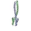
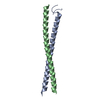
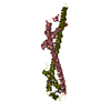
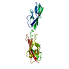
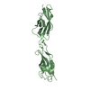
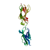
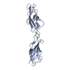
| Title | Crystal Structure of the pre-assembly scaffolding protein gp7 from the double-stranded DNA bacteriophage phi29 | ||||||
 Components Components | HEAD MORPHOGENESIS PROTEIN | ||||||
 Keywords Keywords | VIRAL PROTEIN / coiled-coil | ||||||
| Function / homology |  Function and homology information Function and homology information | ||||||
| Biological species |   Bacillus phage phi29 (virus) Bacillus phage phi29 (virus) | ||||||
| Method |  X-RAY DIFFRACTION / SYNCHROTRON / MAD / Resolution: 2.2 Å X-RAY DIFFRACTION / SYNCHROTRON / MAD / Resolution: 2.2 Å | ||||||
 Authors Authors | Morais, M.C. / Kanamaru, S. / Badasso, M.O. / Koti, J.S. / Owen, B.A.L. / McMurray, C.T. / Anderson, D.L. / Rossmann, M.G. | ||||||
 Citation Citation | Journal: Nat.Struct.Biol. / Year: 2003 Title: Bacteriophage f29 scaffolding protein gp7 before and after prohead assembly Authors: Morais, M.C. / Kanamaru, S. / Badasso, M.O. / Koti, J.S. / Owen, B.A.L. / McMurray, C.T. / Anderson, D.L. / Rossmann, M.G. | ||||||
| History |
|
- Structure visualization
| Structure viewer | Molecule: MolmilJmol/JSmol |
|---|
- Downloads & links
-Download
| PDBx/mmCIF format | 1no4.cif.gz | 72.1 KB | Display | PDBx/mmCIF format |
|---|---|---|---|---|
| PDB format | pdb1no4.ent.gz | 55.4 KB | Display | PDB format |
| PDBx/mmJSON format | 1no4.json.gz | Tree view | PDBx/mmJSON format | |
| Others |  Other downloads Other downloads |
-Validation report
| Arichive directory | https://data.pdbj.org/pub/pdb/validation_reports/no/1no4ftp://data.pdbj.org/pub/pdb/validation_reports/no/1no4 | HTTPS FTP |
|---|
-Related structure data


-Links
 PDBj
PDBj- Assembly
| Deposited unit | 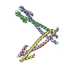
| ||||||||||
|---|---|---|---|---|---|---|---|---|---|---|---|
| 1 | 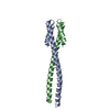
| ||||||||||
| 2 | 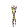
| ||||||||||
| 3 |
| ||||||||||
| Unit cell |
| ||||||||||
| Details | The biological unit is a dimeric coiled-coil. There are two such units per asymmetric unit. |
-Components
| #1: Protein | Mass: 11165.360 Da / Num. of mol.: 4 Source method: isolated from a genetically manipulated source Source: (gene. exp.) Bacillus phage phi29 (virus) / Genus: Phi29-like viruses / Gene: gp7 / Plasmid: pARgp7 / Production host:  #2: Water | ChemComp-HOH / |  Mass: 18.015 Da / Num. of mol.: 129 / Source method: isolated from a natural source / Formula: H2O Mass: 18.015 Da / Num. of mol.: 129 / Source method: isolated from a natural source / Formula: H2O |
|---|
-Experimental details
-Experiment
| Experiment | Method: X-RAY DIFFRACTION / Number of used crystals: 1 |
|---|
- Sample preparation
| Crystal | Density Matthews: 5.56 Å3/Da / Density % sol: 77.7 % | ||||||||||||||||||||||||||||||||||||
|---|---|---|---|---|---|---|---|---|---|---|---|---|---|---|---|---|---|---|---|---|---|---|---|---|---|---|---|---|---|---|---|---|---|---|---|---|---|
| Crystal grow | Temperature: 298 K / Method: vapor diffusion, hanging drop / pH: 6.5 Details: 4 % PEG 8000, 0.16 M calcium acetate, 0.08 M sodium cacodylate, 20 % glycerol, pH 6.5, VAPOR DIFFUSION, HANGING DROP, temperature 298K | ||||||||||||||||||||||||||||||||||||
| Crystal grow | *PLUS Method: unknown | ||||||||||||||||||||||||||||||||||||
| Components of the solutions | *PLUS
|
-Data collection
| Diffraction | Mean temperature: 100 K |
|---|---|
| Diffraction source | Source: SYNCHROTRON / Site: APS  / Beamline: 14-BM-C / Wavelength: 1 Å / Beamline: 14-BM-C / Wavelength: 1 Å |
| Detector | Type: ADSC QUANTUM 4 / Detector: CCD / Date: Nov 17, 2001 |
| Radiation | Protocol: SINGLE WAVELENGTH / Monochromatic (M) / Laue (L): M / Scattering type: x-ray |
| Radiation wavelength | Wavelength: 1 Å / Relative weight: 1 |
| Reflection | Resolution: 2.2→60.3 Å / Num. all: 38534 / Num. obs: 38534 / % possible obs: 95.7 % / Observed criterion σ(F): 0 / Observed criterion σ(I): 0 / Redundancy: 20.06 % / Biso Wilson estimate: 32.8 Å2 / Rsym value: 0.068 / Net I/σ(I): 19.5 |
| Reflection shell | Resolution: 2.2→2.26 Å / Mean I/σ(I) obs: 5 / Num. unique all: 1495 / Rsym value: 0.255 / % possible all: 75.4 |
| Reflection | *PLUS Highest resolution: 2.2 Å / Num. obs: 40285 / Rmerge(I) obs: 0.068 |
| Reflection shell | *PLUS % possible obs: 75.4 % / Rmerge(I) obs: 0.251 |
- Processing
| Software |
| |||||||||||||||||||||||||
|---|---|---|---|---|---|---|---|---|---|---|---|---|---|---|---|---|---|---|---|---|---|---|---|---|---|---|
| Refinement | Method to determine structure: MAD / Resolution: 2.2→60.3 Å / Rfactor Rfree error: 0.004 / Isotropic thermal model: RESTRAINED / Cross valid method: THROUGHOUT / σ(F): 0 / Stereochemistry target values: Engh & Huber
| |||||||||||||||||||||||||
| Solvent computation | Solvent model: FLAT MODEL / Bsol: 58.7035 Å2 / ksol: 0.384725 e/Å3 | |||||||||||||||||||||||||
| Displacement parameters | Biso mean: 58.3 Å2
| |||||||||||||||||||||||||
| Refine analyze |
| |||||||||||||||||||||||||
| Refinement step | Cycle: LAST / Resolution: 2.2→60.3 Å
| |||||||||||||||||||||||||
| Refine LS restraints |
| |||||||||||||||||||||||||
| LS refinement shell | Resolution: 2.2→2.34 Å / Rfactor Rfree error: 0.015 / Total num. of bins used: 6
| |||||||||||||||||||||||||
| Xplor file |
| |||||||||||||||||||||||||
| Refinement | *PLUS Highest resolution: 2.2 Å | |||||||||||||||||||||||||
| Solvent computation | *PLUS | |||||||||||||||||||||||||
| Displacement parameters | *PLUS | |||||||||||||||||||||||||
| Refine LS restraints | *PLUS
|
