 Movie
Movie Controller

[English] 日本語
 Yorodumi
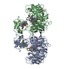
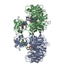
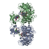
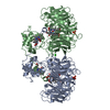
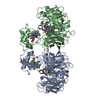
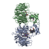
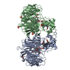
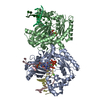
- PDB-1nno: CONFORMATIONAL CHANGES OCCURRING UPON NO BINDING IN NITRITE REDUC... -
+ Open data

- Basic information
| Entry | Database: PDB / ID: 1nno | ||||||
|---|---|---|---|---|---|---|---|
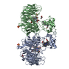
| Title | CONFORMATIONAL CHANGES OCCURRING UPON NO BINDING IN NITRITE REDUCTASE FROM PSEUDOMONAS AERUGINOSA | ||||||
 Components Components | NITRITE REDUCTASE | ||||||
 Keywords Keywords | OXIDOREDUCTASE / NITRITE REDUCTASE / PSEUDOMONAS AERUGINOSA / HEMOPROTEIN / DENITRIFICATION / NO BINDING / CONFORMATIONAL CHANGES | ||||||
| Function / homology |  Function and homology information Function and homology informationhydroxylamine reductase / hydroxylamine reductase activity / nitrite reductase (NO-forming) / nitrite reductase (NO-forming) activity / electron transfer activity / periplasmic space / heme binding / metal ion binding Similarity search - Function | ||||||
| Biological species |   Pseudomonas aeruginosa (bacteria) Pseudomonas aeruginosa (bacteria) | ||||||
| Method |  X-RAY DIFFRACTION / SYNCHROTRON / OTHER / Resolution: 2.65 Å X-RAY DIFFRACTION / SYNCHROTRON / OTHER / Resolution: 2.65 Å | ||||||
 Authors Authors | Nurizzo, D. / Tegoni, M. / Cambillau, C. | ||||||
 Citation Citation | Journal: Biochemistry / Year: 1998 Title: Conformational changes occurring upon reduction and NO binding in nitrite reductase from Pseudomonas aeruginosa. Authors: Nurizzo, D. / Cutruzzola, F. / Arese, M. / Bourgeois, D. / Brunori, M. / Cambillau, C. / Tegoni, M. | ||||||
| History |
|
- Structure visualization
| Structure viewer | Molecule: MolmilJmol/JSmol |
|---|
- Downloads & links
-Download
| PDBx/mmCIF format | 1nno.cif.gz | 212.8 KB | Display | PDBx/mmCIF format |
|---|---|---|---|---|
| PDB format | pdb1nno.ent.gz | 171.8 KB | Display | PDB format |
| PDBx/mmJSON format | 1nno.json.gz | Tree view | PDBx/mmJSON format | |
| Others |  Other downloads Other downloads |
-Validation report
| Arichive directory | https://data.pdbj.org/pub/pdb/validation_reports/nn/1nnoftp://data.pdbj.org/pub/pdb/validation_reports/nn/1nno | HTTPS FTP |
|---|
-Related structure data


-Links
 PDBj
PDBj


- Assembly
| Deposited unit | 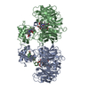
| ||||||||
|---|---|---|---|---|---|---|---|---|---|
| 1 |
| ||||||||
| Unit cell |
| ||||||||
| Noncrystallographic symmetry (NCS) | NCS oper: (Code: given Matrix: (0.999943, -0.001407, -0.010572), Vector: |
-Components
| #1: Protein | Mass: 60259.051 Da / Num. of mol.: 2 / Source method: isolated from a natural source Details: EACH SUBUNIT (A,B) LINKS ONE NITRIC OXIDE (NO) AT THE CATALYTIC SITE Source: (natural) Pseudomonas aeruginosa (bacteria) / Cellular location: PERIPLASMIC SPACE / References: UniProt: P24474, EC: 1.9.3.2#2: Chemical |   Mass: 618.503 Da / Num. of mol.: 2 / Source method: obtained synthetically / Formula: C34H34FeN4O4 Mass: 618.503 Da / Num. of mol.: 2 / Source method: obtained synthetically / Formula: C34H34FeN4O4#3: Chemical | 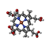  Mass: 712.484 Da / Num. of mol.: 2 / Source method: obtained synthetically / Formula: C34H32FeN4O10 Mass: 712.484 Da / Num. of mol.: 2 / Source method: obtained synthetically / Formula: C34H32FeN4O10#4: Chemical |   Mass: 30.006 Da / Num. of mol.: 2 / Source method: obtained synthetically / Formula: NO Mass: 30.006 Da / Num. of mol.: 2 / Source method: obtained synthetically / Formula: NO#5: Water | ChemComp-HOH / |  Mass: 18.015 Da / Num. of mol.: 354 / Source method: isolated from a natural source / Formula: H2O Mass: 18.015 Da / Num. of mol.: 354 / Source method: isolated from a natural source / Formula: H2OHas protein modification | Y | |
|---|
-Experimental details
-Experiment
| Experiment | Method: X-RAY DIFFRACTION / Number of used crystals: 1 |
|---|
- Sample preparation
| Crystal | Density Matthews: 3.47 Å3/Da / Density % sol: 64.6 % | |||||||||||||||
|---|---|---|---|---|---|---|---|---|---|---|---|---|---|---|---|---|
| Crystal grow | pH: 7.2 / Details: pH 7.2 | |||||||||||||||
| Crystal | *PLUS | |||||||||||||||
| Crystal grow | *PLUS pH: 8.4 / Method: unknown | |||||||||||||||
| Components of the solutions | *PLUS
|
-Data collection
| Diffraction | Mean temperature: 100 K |
|---|---|
| Diffraction source | Source: SYNCHROTRON / Site: EMBL/DESY, HAMBURG  / Beamline: X11 / Wavelength: 0.907 / Beamline: X11 / Wavelength: 0.907 |
| Detector | Type: MARRESEARCH / Detector: IMAGE PLATE / Date: Dec 1, 1996 |
| Radiation | Monochromatic (M) / Laue (L): M / Scattering type: x-ray |
| Radiation wavelength | Wavelength: 0.907 Å / Relative weight: 1 |
| Reflection | Resolution: 2.65→15 Å / Num. obs: 44045 / % possible obs: 90.7 % / Observed criterion σ(I): 2 / Redundancy: 3 % / Rmerge(I) obs: 0.069 / Net I/σ(I): 10.3 |
| Reflection shell | Resolution: 2.65→2.71 Å / Rmerge(I) obs: 0.479 / Mean I/σ(I) obs: 2.1 / % possible all: 93.3 |
| Reflection | *PLUS Num. measured all: 361716 / Rmerge(I) obs: 0.11 |
| Reflection shell | *PLUS % possible obs: 93.3 % / Rmerge(I) obs: 0.569 |
- Processing
| Software |
| ||||||||||||||||||||||||||||||||||||||||||||||||||||||||||||
|---|---|---|---|---|---|---|---|---|---|---|---|---|---|---|---|---|---|---|---|---|---|---|---|---|---|---|---|---|---|---|---|---|---|---|---|---|---|---|---|---|---|---|---|---|---|---|---|---|---|---|---|---|---|---|---|---|---|---|---|---|---|
| Refinement | Method to determine structure: OTHER / Resolution: 2.65→15 Å / Rfactor Rfree error: 0.0052 / Data cutoff high absF: 100000 / Data cutoff low absF: 0.01 / Cross valid method: THROUGHOUT / σ(F): 1
| ||||||||||||||||||||||||||||||||||||||||||||||||||||||||||||
| Displacement parameters |
| ||||||||||||||||||||||||||||||||||||||||||||||||||||||||||||
| Refine analyze | Luzzati d res low obs: 15 Å | ||||||||||||||||||||||||||||||||||||||||||||||||||||||||||||
| Refinement step | Cycle: LAST / Resolution: 2.65→15 Å
| ||||||||||||||||||||||||||||||||||||||||||||||||||||||||||||
| Refine LS restraints |
| ||||||||||||||||||||||||||||||||||||||||||||||||||||||||||||
| LS refinement shell | Resolution: 2.65→2.77 Å / Rfactor Rfree error: 0.024 / Total num. of bins used: 8
| ||||||||||||||||||||||||||||||||||||||||||||||||||||||||||||
| Xplor file |
| ||||||||||||||||||||||||||||||||||||||||||||||||||||||||||||
| Software | *PLUS Name: X-PLOR / Version: 3.843 / Classification: refinement | ||||||||||||||||||||||||||||||||||||||||||||||||||||||||||||
| Refinement | *PLUS Rfactor Rfree: 0.2438 | ||||||||||||||||||||||||||||||||||||||||||||||||||||||||||||
| Solvent computation | *PLUS | ||||||||||||||||||||||||||||||||||||||||||||||||||||||||||||
| Displacement parameters | *PLUS | ||||||||||||||||||||||||||||||||||||||||||||||||||||||||||||
| Refine LS restraints | *PLUS
|
