 Movie
Movie Controller


+ Open data

- Basic information
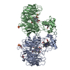
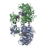
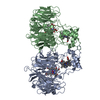
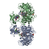
| Entry | Database: PDB / ID: 1gq1 | ||||||
|---|---|---|---|---|---|---|---|
| Title | CYTOCHROME CD1 NITRITE REDUCTASE, Y25S mutant, OXIDISED FORM | ||||||
 Components Components | CYTOCHROME CD1 NITRITE REDUCTASE | ||||||
 Keywords Keywords | OXIDOREDUCTASE / REDUCTASE / ENZYME / NITRITE REDUCTASE / DENITRIFICATION / ELECTRON TRANSPORT / PERIPLASMIC | ||||||
| Function / homology |  Function and homology information Function and homology informationhydroxylamine reductase / hydroxylamine reductase activity / nitrite reductase (NO-forming) / nitrite reductase (NO-forming) activity / electron transfer activity / periplasmic space / heme binding / metal ion binding Similarity search - Function | ||||||
| Biological species |  PARACOCCUS PANTOTROPHUS (bacteria) PARACOCCUS PANTOTROPHUS (bacteria) | ||||||
| Method |  X-RAY DIFFRACTION / SYNCHROTRON / MOLECULAR REPLACEMENT / Resolution: 1.4 Å X-RAY DIFFRACTION / SYNCHROTRON / MOLECULAR REPLACEMENT / Resolution: 1.4 Å | ||||||
 Authors Authors | Sjogren, T. / Gordon, E.H.J. / Lofqvist, M. / Richter, C.D. / Hajdu, J. / Ferguson, S.J. | ||||||
 Citation Citation | Journal: J.Biol.Chem. / Year: 2003 Title: Structure and Kinetic Properties of Paracoccus Pantotrophus Cytochrome Cd1 Nitrite Reductase with the D1 Heme Active Site Ligand Tyrosine 25 Replaced by Serine Authors: Gordon, E.H.J. / Sjogren, T. / Lofqvist, M. / Richter, C.D. / Allen, J. / Higham, C. / Hajdu, J. / Fulop, V. / Ferguson, S.J. #1: Journal: Cell(Cambridge,Mass.) / Year: 1995Title: The Anatomy of a Bifunctional Enzyme: Structural Basis for Reduction of Oxygen to Water and Synthesis of Nitric Oxide by Cytochrome Cd1 Authors: Fulop, V. / Moir, J.W.B. / Ferguson, S.J. / Hajdu, J. | ||||||
| History |
|
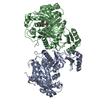
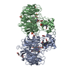
- Structure visualization
| Structure viewer | Molecule: MolmilJmol/JSmol |
|---|
- Downloads & links
-Download
| PDBx/mmCIF format | 1gq1.cif.gz | 262.7 KB | Display | PDBx/mmCIF format |
|---|---|---|---|---|
| PDB format | pdb1gq1.ent.gz | 209.7 KB | Display | PDB format |
| PDBx/mmJSON format | 1gq1.json.gz | Tree view | PDBx/mmJSON format | |
| Others |  Other downloads Other downloads |
-Validation report
| Arichive directory | https://data.pdbj.org/pub/pdb/validation_reports/gq/1gq1ftp://data.pdbj.org/pub/pdb/validation_reports/gq/1gq1 | HTTPS FTP |
|---|
-Related structure data
| Related structure data |  1qksS S: Starting model for refinement |
|---|---|
| Similar structure data |


-Links
 PDBj
PDBj


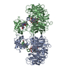
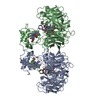
- Assembly
| Deposited unit | 
| ||||||||
|---|---|---|---|---|---|---|---|---|---|
| 1 |
| ||||||||
| Unit cell |
|
-Components
-Protein , 1 types, 2 molecules AB
| #1: Protein | Mass: 62470.438 Da / Num. of mol.: 2 / Mutation: YES Source method: isolated from a genetically manipulated source Source: (gene. exp.) PARACOCCUS PANTOTROPHUS (bacteria) / Plasmid: PEG276 / Production host: PARACOCCUS PANTOTROPHUS (bacteria) / Strain (production host): EG6202References: UniProt: Q9FCQ0, UniProt: P72181*PLUS, EC: 1.9.3.2, nitrite reductase (NO-forming) |
|---|
-Non-polymers , 5 types, 1369 molecules 
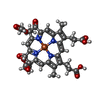


| #2: Chemical |  Mass: 618.503 Da / Num. of mol.: 2 / Source method: obtained synthetically / Formula: C34H34FeN4O4 Mass: 618.503 Da / Num. of mol.: 2 / Source method: obtained synthetically / Formula: C34H34FeN4O4#3: Chemical |  Mass: 712.484 Da / Num. of mol.: 2 / Source method: obtained synthetically / Formula: C34H32FeN4O10 Mass: 712.484 Da / Num. of mol.: 2 / Source method: obtained synthetically / Formula: C34H32FeN4O10#4: Chemical | ChemComp-SO4 /  Mass: 96.063 Da / Num. of mol.: 8 / Source method: obtained synthetically / Formula: SO4 Mass: 96.063 Da / Num. of mol.: 8 / Source method: obtained synthetically / Formula: SO4#5: Chemical |  Mass: 92.094 Da / Num. of mol.: 3 / Source method: obtained synthetically / Formula: C3H8O3 Mass: 92.094 Da / Num. of mol.: 3 / Source method: obtained synthetically / Formula: C3H8O3#6: Water | ChemComp-HOH / | Mass: 18.015 Da / Num. of mol.: 1354 / Source method: isolated from a natural source / Formula: H2O |
|---|
-Details
| Compound details | ENGINEERED| Has protein modification | Y | |
|---|
-Experimental details
-Experiment
| Experiment | Method: X-RAY DIFFRACTION / Number of used crystals: 1 |
|---|
- Sample preparation
| Crystal | Density Matthews: 2.52 Å3/Da / Density % sol: 51 % | ||||||||||||||||||||||||||||||
|---|---|---|---|---|---|---|---|---|---|---|---|---|---|---|---|---|---|---|---|---|---|---|---|---|---|---|---|---|---|---|---|
| Crystal grow | pH: 7 Details: 2.3 M AMMONIUM SULFATE, 50MM POTASSIUM PHOSPHATE, PH 7.0, AND CRYOPROTECTANT 18% GLYCEROL | ||||||||||||||||||||||||||||||
| Crystal grow | *PLUS Temperature: 15 ℃ / Method: vapor diffusion, hanging drop / Details: Fulop, V., (1993) J. Mol. Biol., 232, 1211. | ||||||||||||||||||||||||||||||
| Components of the solutions | *PLUS
|
-Data collection
| Diffraction | Mean temperature: 100 K |
|---|---|
| Diffraction source | Source: SYNCHROTRON / Site: MAX II  / Beamline: I711 / Wavelength: 0.934 / Beamline: I711 / Wavelength: 0.934 |
| Detector | Type: MARRESEARCH / Detector: IMAGE PLATE / Date: Oct 6, 2000 / Details: MIRRORS |
| Radiation | Monochromator: SI CRYSTAL / Protocol: SINGLE WAVELENGTH / Monochromatic (M) / Laue (L): M / Scattering type: x-ray |
| Radiation wavelength | Wavelength: 0.934 Å / Relative weight: 1 |
| Reflection | Resolution: 1.4→30 Å / Num. obs: 233906 / % possible obs: 100 % / Observed criterion σ(I): -3 / Redundancy: 3.3 % / Rmerge(I) obs: 0.048 / Net I/σ(I): 24.8 |
| Reflection shell | Resolution: 1.4→1.45 Å / Redundancy: 3.1 % / Rmerge(I) obs: 0.247 / Mean I/σ(I) obs: 4.3 / % possible all: 100 |
| Reflection | *PLUS Lowest resolution: 30 Å / % possible obs: 100 % / Num. measured all: 812906 |
| Reflection shell | *PLUS Highest resolution: 1.4 Å / % possible obs: 100 % / Mean I/σ(I) obs: 4.72 |
- Processing
| Software |
| ||||||||||||||||||||
|---|---|---|---|---|---|---|---|---|---|---|---|---|---|---|---|---|---|---|---|---|---|
| Refinement | Method to determine structure: MOLECULAR REPLACEMENT Starting model: PDB ENTRY 1QKS Resolution: 1.4→30 Å / SU B: 0.79 / SU ML: 0.03 / Cross valid method: THROUGHOUT / σ(F): 2 / ESU R: 0.05 / ESU R Free: 0.05
| ||||||||||||||||||||
| Displacement parameters | Biso mean: 10.9 Å2 | ||||||||||||||||||||
| Refinement step | Cycle: LAST / Resolution: 1.4→30 Å
| ||||||||||||||||||||
| Refinement | *PLUS Lowest resolution: 30 Å / % reflection Rfree: 4 % | ||||||||||||||||||||
| Solvent computation | *PLUS | ||||||||||||||||||||
| Displacement parameters | *PLUS | ||||||||||||||||||||
| Refine LS restraints | *PLUS
|
