 Movie
Movie Controller


+ Open data

- Basic information
| Entry | Database: PDB / ID: 1nmp | ||||||
|---|---|---|---|---|---|---|---|
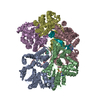
| Title | Structural genomics, ybgI protein, unknown function | ||||||
 Components Components | Hypothetical protein ybgI | ||||||
 Keywords Keywords | structural genomics / unknown function / ybgI / hypothetical protein / toroidal structure / Structure 2 Function Project / S2F | ||||||
| Function / homology |  Function and homology information Function and homology informationcell pole / protein hexamerization / response to ionizing radiation / DNA repair / metal ion binding / identical protein binding / cytosol / cytoplasm Similarity search - Function | ||||||
| Biological species |  | ||||||
| Method |  X-RAY DIFFRACTION / SYNCHROTRON / MOLECULAR REPLACEMENT / Resolution: 2.2 Å X-RAY DIFFRACTION / SYNCHROTRON / MOLECULAR REPLACEMENT / Resolution: 2.2 Å | ||||||
 Authors Authors | Ladner, J.E. / Obmolova, G. / Teplyakov, A. / Khil, P.P. / Camerini-Otero, R.D. / Gilliland, G.L. / Structure 2 Function Project (S2F) | ||||||
 Citation Citation | Journal: BMC Struct.Biol. / Year: 2003 Title: Crystal Structure of Escherichia coli Protein ybgI, a toroidal structure with a dinuclear metal site Authors: Ladner, J.E. / Obmolova, G. / Teplyakov, A. / Howard, A.J. / Khil, P.P. / Camerini-Otero, R.D. / Gilliland, G.L. | ||||||
| History |
| ||||||
| Remark 600 | HETEROGEN THE IDENTIFICATION OF THE METAL ION IN THE ACTIVE SITE IS NOT KNOWN. HERE IT IS MODELED AS MG+2. |
- Structure visualization
| Structure viewer | Molecule: MolmilJmol/JSmol |
|---|
- Downloads & links
-Download
| PDBx/mmCIF format | 1nmp.cif.gz | 294.2 KB | Display | PDBx/mmCIF format |
|---|---|---|---|---|
| PDB format | pdb1nmp.ent.gz | 239.6 KB | Display | PDB format |
| PDBx/mmJSON format | 1nmp.json.gz | Tree view | PDBx/mmJSON format | |
| Others |  Other downloads Other downloads |
-Validation report
| Arichive directory | https://data.pdbj.org/pub/pdb/validation_reports/nm/1nmpftp://data.pdbj.org/pub/pdb/validation_reports/nm/1nmp | HTTPS FTP |
|---|
-Related structure data

-Links
 PDBj
PDBj- Assembly
| Deposited unit | 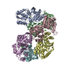
| ||||||||
|---|---|---|---|---|---|---|---|---|---|
| 1 |
| ||||||||
| 2 | 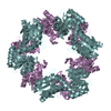
| ||||||||
| 3 | 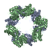
| ||||||||
| 4 | 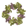
| ||||||||
| Unit cell |
| ||||||||
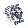
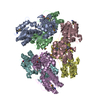
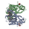
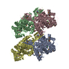
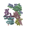
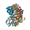
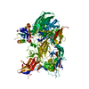
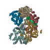
| Details | The biological assembly is a hexamer generated by the application of the symmetry operations: -y, x-y, Z and y-x, -x, z. If applied to the entire asymmetric unit, these operations will generate 3 toroids (3 biological assemblies). If applied to any of the dimers, AB, CD or EF, these operations will produce one biological assembly. |
-Components
| #1: Protein | Mass: 26919.400 Da / Num. of mol.: 6 Source method: isolated from a genetically manipulated source Source: (gene. exp.) Escherichia coli, Escherichia coli O157:H7 Genus: Escherichia, Escherichia / Species: , Escherichia coli / Strain: , O157:H7 / Gene: YBGI OR B0710 OR Z0861 OR ECS0735 / Plasmid: pDEST14 / Production host: #2: Chemical | ChemComp-MG /   Mass: 24.305 Da / Num. of mol.: 11 / Source method: obtained synthetically / Formula: Mg Mass: 24.305 Da / Num. of mol.: 11 / Source method: obtained synthetically / Formula: Mg#3: Water | ChemComp-HOH / |  Mass: 18.015 Da / Num. of mol.: 576 / Source method: isolated from a natural source / Formula: H2O Mass: 18.015 Da / Num. of mol.: 576 / Source method: isolated from a natural source / Formula: H2O |
|---|
-Experimental details
-Experiment
| Experiment | Method: X-RAY DIFFRACTION / Number of used crystals: 1 |
|---|
- Sample preparation
| Crystal | Density Matthews: 2.44 Å3/Da / Density % sol: 49.15 % | ||||||||||||||||||||||||||||||||||||
|---|---|---|---|---|---|---|---|---|---|---|---|---|---|---|---|---|---|---|---|---|---|---|---|---|---|---|---|---|---|---|---|---|---|---|---|---|---|
| Crystal grow | Temperature: 298 K / Method: vapor diffusion, hanging drop / pH: 7.5 Details: 0.1M cacodylate buffer pH 7.5, 0.1M magnesium acetate, 15% (w/v) polyethylene glycol 8000, 5% (v/v) polyethylene glycol 400, VAPOR DIFFUSION, HANGING DROP, temperature 298K | ||||||||||||||||||||||||||||||||||||
| Crystal grow | *PLUS Method: vapor diffusion, hanging drop | ||||||||||||||||||||||||||||||||||||
| Components of the solutions | *PLUS
|
-Data collection
| Diffraction | Mean temperature: 100 K |
|---|---|
| Diffraction source | Source: SYNCHROTRON / Site: APS  / Beamline: 22-ID / Wavelength: 0.9793 Å / Beamline: 22-ID / Wavelength: 0.9793 Å |
| Detector | Type: MARRESEARCH / Detector: CCD / Date: Jul 2, 2002 |
| Radiation | Protocol: SINGLE WAVELENGTH / Monochromatic (M) / Laue (L): M / Scattering type: x-ray |
| Radiation wavelength | Wavelength: 0.9793 Å / Relative weight: 1 |
| Reflection | Resolution: 2.2→20 Å / Num. obs: 80094 / Redundancy: 5 % / Biso Wilson estimate: 4.1 Å2 / Rmerge(I) obs: 0.096 / Net I/σ(I): 14.1 |
| Reflection | *PLUS % possible obs: 99.8 % / Num. measured all: 303926 |
| Reflection shell | *PLUS % possible obs: 99.6 % / Rmerge(I) obs: 0.237 / Mean I/σ(I) obs: 2.8 |
- Processing
| Software |
| ||||||||||||||||||||||||||||||||||||||||||||||||||||||||||||||||||||||||||||||||
|---|---|---|---|---|---|---|---|---|---|---|---|---|---|---|---|---|---|---|---|---|---|---|---|---|---|---|---|---|---|---|---|---|---|---|---|---|---|---|---|---|---|---|---|---|---|---|---|---|---|---|---|---|---|---|---|---|---|---|---|---|---|---|---|---|---|---|---|---|---|---|---|---|---|---|---|---|---|---|---|---|---|
| Refinement | Method to determine structure: MOLECULAR REPLACEMENT Starting model: SeMet structure Resolution: 2.2→19.76 Å / Rfactor Rfree error: 0.003 / Data cutoff high absF: 199707.79 / Data cutoff high rms absF: 199707.79 / Data cutoff low absF: 0 / Isotropic thermal model: RESTRAINED / Cross valid method: THROUGHOUT / σ(F): 0
| ||||||||||||||||||||||||||||||||||||||||||||||||||||||||||||||||||||||||||||||||
| Solvent computation | Solvent model: FLAT MODEL / Bsol: 26.9411 Å2 / ksol: 0.352375 e/Å3 | ||||||||||||||||||||||||||||||||||||||||||||||||||||||||||||||||||||||||||||||||
| Displacement parameters | Biso mean: 13.6 Å2
| ||||||||||||||||||||||||||||||||||||||||||||||||||||||||||||||||||||||||||||||||
| Refine analyze | Luzzati coordinate error free: 0.32 Å / Luzzati sigma a free: 0.25 Å | ||||||||||||||||||||||||||||||||||||||||||||||||||||||||||||||||||||||||||||||||
| Refinement step | Cycle: LAST / Resolution: 2.2→19.76 Å
| ||||||||||||||||||||||||||||||||||||||||||||||||||||||||||||||||||||||||||||||||
| Refine LS restraints |
| ||||||||||||||||||||||||||||||||||||||||||||||||||||||||||||||||||||||||||||||||
| Refine LS restraints NCS | NCS model details: CONSTR | ||||||||||||||||||||||||||||||||||||||||||||||||||||||||||||||||||||||||||||||||
| LS refinement shell | Resolution: 2.2→2.34 Å / Rfactor Rfree error: 0.008 / Total num. of bins used: 6
| ||||||||||||||||||||||||||||||||||||||||||||||||||||||||||||||||||||||||||||||||
| Xplor file |
| ||||||||||||||||||||||||||||||||||||||||||||||||||||||||||||||||||||||||||||||||
| Refinement | *PLUS Highest resolution: 2.2 Å / Lowest resolution: 20 Å / % reflection Rfree: 5 % / Rfactor Rfree: 0.252 / Rfactor Rwork: 0.213 | ||||||||||||||||||||||||||||||||||||||||||||||||||||||||||||||||||||||||||||||||
| Solvent computation | *PLUS | ||||||||||||||||||||||||||||||||||||||||||||||||||||||||||||||||||||||||||||||||
| Displacement parameters | *PLUS | ||||||||||||||||||||||||||||||||||||||||||||||||||||||||||||||||||||||||||||||||
| Refine LS restraints | *PLUS
|
