 Movie
Movie Controller


+ Open data

- Basic information
| Entry | Database: PDB / ID: 1nks | ||||||
|---|---|---|---|---|---|---|---|
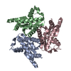
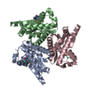
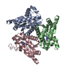
| Title | ADENYLATE KINASE FROM SULFOLOBUS ACIDOCALDARIUS | ||||||
 Components Components | ADENYLATE KINASE | ||||||
 Keywords Keywords | KINASE / THERMOPHILIC / TRANSFERASE | ||||||
| Function / homology |  Function and homology information Function and homology information | ||||||
| Biological species |   Sulfolobus acidocaldarius (acidophilic) Sulfolobus acidocaldarius (acidophilic) | ||||||
| Method |  X-RAY DIFFRACTION / SYNCHROTRON / MIR / Resolution: 2.57 Å X-RAY DIFFRACTION / SYNCHROTRON / MIR / Resolution: 2.57 Å | ||||||
 Authors Authors | Vonrhein, C. / Schulz, G.E. | ||||||
 Citation Citation | Journal: J.Mol.Biol. / Year: 1998 Title: The structure of a trimeric archaeal adenylate kinase. Authors: Vonrhein, C. / Bonisch, H. / Schafer, G. / Schulz, G.E. | ||||||
| History |
|
- Structure visualization
| Structure viewer | Molecule: MolmilJmol/JSmol |
|---|
- Downloads & links
-Download
| PDBx/mmCIF format | 1nks.cif.gz | 236.5 KB | Display | PDBx/mmCIF format |
|---|---|---|---|---|
| PDB format | pdb1nks.ent.gz | 193.7 KB | Display | PDB format |
| PDBx/mmJSON format | 1nks.json.gz | Tree view | PDBx/mmJSON format | |
| Others |  Other downloads Other downloads |
-Validation report
| Arichive directory | https://data.pdbj.org/pub/pdb/validation_reports/nk/1nksftp://data.pdbj.org/pub/pdb/validation_reports/nk/1nks | HTTPS FTP |
|---|
-Related structure data
| Similar structure data |
|---|

-Links
 PDBj
PDBj


- Assembly
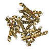
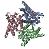
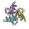
| Deposited unit | 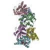
| ||||||||
|---|---|---|---|---|---|---|---|---|---|
| 1 | 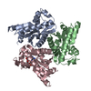
| ||||||||
| 2 | 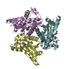
| ||||||||
| 3 | 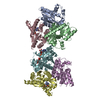
| ||||||||
| 4 | 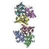
| ||||||||
| Unit cell |
|
-Components
| #1: Protein | Mass: 21137.369 Da / Num. of mol.: 6 Source method: isolated from a genetically manipulated source Source: (gene. exp.) Sulfolobus acidocaldarius (acidophilic)Plasmid: PET3A / Cell line (production host): BL21 (DE3) / Production host:  #2: Chemical |   Mass: 347.221 Da / Num. of mol.: 3 / Source method: obtained synthetically / Formula: C10H14N5O7P / Comment: AMP*YM Mass: 347.221 Da / Num. of mol.: 3 / Source method: obtained synthetically / Formula: C10H14N5O7P / Comment: AMP*YM#3: Chemical | ChemComp-ADP / |   Mass: 427.201 Da / Num. of mol.: 1 / Source method: obtained synthetically / Formula: C10H15N5O10P2 / Comment: ADP, energy-carrying molecule*YM Mass: 427.201 Da / Num. of mol.: 1 / Source method: obtained synthetically / Formula: C10H15N5O10P2 / Comment: ADP, energy-carrying molecule*YM#4: Water | ChemComp-HOH / |  Mass: 18.015 Da / Num. of mol.: 332 / Source method: isolated from a natural source / Formula: H2O Mass: 18.015 Da / Num. of mol.: 332 / Source method: isolated from a natural source / Formula: H2O |
|---|
-Experimental details
-Experiment
| Experiment | Method: X-RAY DIFFRACTION / Number of used crystals: 4 |
|---|
- Sample preparation
| Crystal | Density Matthews: 3.63 Å3/Da / Density % sol: 66 % Description: LOW RESOLUTION DATA WERE COLLECTED ON A ROTATING ANODE AND MERGED WITH HIGH-RESOLUTION DATA COLLECTED FROM THE SYNCHROTRON SOURCE DESCRIBED ABOVE. | ||||||||||||||||||||
|---|---|---|---|---|---|---|---|---|---|---|---|---|---|---|---|---|---|---|---|---|---|
| Crystal grow | pH: 7.5 Details: PROTEIN WAS CRYSTALLIZED FROM 2 M SODIUM FORMATE AT PH 7.5 AT A PROTEIN CONCENTRATION OF 5 MG/ML; PROTEIN SOLUTION WAS QUARTZ-DISTILLED AND NO NUCLEOTIDES WERE ADDED. | ||||||||||||||||||||
| Crystal grow | *PLUS Method: vapor diffusion, hanging drop | ||||||||||||||||||||
| Components of the solutions | *PLUS
|
-Data collection
| Diffraction | Mean temperature: 292 K |
|---|---|
| Diffraction source | Source: SYNCHROTRON / Site: EMBL/DESY, HAMBURG  / Beamline: X11 / Wavelength: 0.9123 / Beamline: X11 / Wavelength: 0.9123 |
| Detector | Type: MARRESEARCH / Detector: IMAGE PLATE / Date: Jun 1, 1996 / Details: DUAL SLITS |
| Radiation | Monochromator: SI(111) / Monochromatic (M) / Laue (L): M / Scattering type: x-ray |
| Radiation wavelength | Wavelength: 0.9123 Å / Relative weight: 1 |
| Reflection | Resolution: 2.57→40 Å / Num. obs: 54962 / % possible obs: 92 % / Observed criterion σ(I): 0 / Redundancy: 3 % / Rsym value: 0.091 / Net I/σ(I): 13.3 |
| Reflection shell | Resolution: 2.57→2.64 Å / Redundancy: 1.4 % / Mean I/σ(I) obs: 2 / Rsym value: 0.266 / % possible all: 56 |
| Reflection | *PLUS Num. measured all: 168355 / Rmerge(I) obs: 0.091 |
| Reflection shell | *PLUS % possible obs: 56 % / Rmerge(I) obs: 0.266 |
- Processing
| Software |
| |||||||||||||||||||||||||||
|---|---|---|---|---|---|---|---|---|---|---|---|---|---|---|---|---|---|---|---|---|---|---|---|---|---|---|---|---|
| Refinement | Method to determine structure: MIR / Resolution: 2.57→40 Å / Cross valid method: THROUGHOUT / σ(F): 0 Details: BULK SOLVENT CORRECTION AS IMPLEMENTED IN X-PLOR 3.851 WAS USED.
| |||||||||||||||||||||||||||
| Refinement step | Cycle: LAST / Resolution: 2.57→40 Å
| |||||||||||||||||||||||||||
| Software | *PLUS Name: REFMAC / Classification: refinement | |||||||||||||||||||||||||||
| Refinement | *PLUS Rfactor obs: 0.164 | |||||||||||||||||||||||||||
| Solvent computation | *PLUS | |||||||||||||||||||||||||||
| Displacement parameters | *PLUS |
