 Movie
Movie Controller

[English] 日本語
 Yorodumi
- PDB-1nb2: Crystal Structure of Nucleoside Diphosphate Kinase from Bacillus ... -
+ Open data

- Basic information
| Entry | Database: PDB / ID: 1nb2 | ||||||
|---|---|---|---|---|---|---|---|
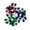
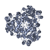
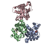
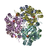
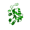
| Title | Crystal Structure of Nucleoside Diphosphate Kinase from Bacillus Halodenitrificans | ||||||
 Components Components | Nucleoside Diphosphate Kinase | ||||||
 Keywords Keywords | TRANSFERASE / Nucleoside Diphosphate Kinase / Bacillus Halodenitrifians | ||||||
| Function / homology |  Function and homology information Function and homology informationnucleoside-diphosphate kinase / UTP biosynthetic process / CTP biosynthetic process / nucleoside diphosphate kinase activity / GTP biosynthetic process / ATP binding / metal ion binding / cytoplasm Similarity search - Function | ||||||
| Biological species |  Virgibacillus halodenitrificans (bacteria) Virgibacillus halodenitrificans (bacteria) | ||||||
| Method |  X-RAY DIFFRACTION / SYNCHROTRON / MOLECULAR REPLACEMENT / Resolution: 2.2 Å X-RAY DIFFRACTION / SYNCHROTRON / MOLECULAR REPLACEMENT / Resolution: 2.2 Å | ||||||
 Authors Authors | Chen, C.-J. / Liu, M.-Y. / Chang, W.-C. / Chang, T. / Wang, B.-C. / Le Gall, J. | ||||||
 Citation Citation | Journal: J.Struct.Biol. / Year: 2003 Title: Crystal structure of a nucleoside diphosphate kinase from Bacillus halodenitrificans: coexpression of its activity with a Mn-superoxide dismutase. Authors: Chen, C.-J. / Liu, M.-Y. / Chang, T. / Chang, W.-C. / Wang, B.-C. / Le Gall, J. | ||||||
| History |
|
- Structure visualization
| Structure viewer | Molecule: MolmilJmol/JSmol |
|---|
- Downloads & links
-Download
| PDBx/mmCIF format | 1nb2.cif.gz | 37.7 KB | Display | PDBx/mmCIF format |
|---|---|---|---|---|
| PDB format | pdb1nb2.ent.gz | 26.8 KB | Display | PDB format |
| PDBx/mmJSON format | 1nb2.json.gz | Tree view | PDBx/mmJSON format | |
| Others |  Other downloads Other downloads |
-Validation report
| Arichive directory | https://data.pdbj.org/pub/pdb/validation_reports/nb/1nb2ftp://data.pdbj.org/pub/pdb/validation_reports/nb/1nb2 | HTTPS FTP |
|---|
-Related structure data
| Similar structure data |
|---|

-Links
 PDBj
PDBj- Assembly
| Deposited unit | 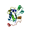
| ||||||||
|---|---|---|---|---|---|---|---|---|---|
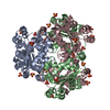
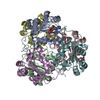
| 1 | x 6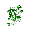
| ||||||||
| Unit cell |
| ||||||||
| Details | The biological assembly is a hexamer generated from the monomer in the asymmetric unit by the crystallographic operations. |
-Components
| #1: Protein | Mass: 14883.729 Da / Num. of mol.: 1 / Source method: isolated from a natural source Source: (natural) Virgibacillus halodenitrificans (bacteria)References: UniProt: Q7SIA9, nucleoside-diphosphate kinase |
|---|---|
| #2: Water | ChemComp-HOH /  Mass: 18.015 Da / Num. of mol.: 34 / Source method: isolated from a natural source / Formula: H2O Mass: 18.015 Da / Num. of mol.: 34 / Source method: isolated from a natural source / Formula: H2O |
-Experimental details
-Experiment
| Experiment | Method: X-RAY DIFFRACTION / Number of used crystals: 1 |
|---|
- Sample preparation
| Crystal | Density Matthews: 4.15 Å3/Da / Density % sol: 70.39 % | ||||||||||||||||||||||||
|---|---|---|---|---|---|---|---|---|---|---|---|---|---|---|---|---|---|---|---|---|---|---|---|---|---|
| Crystal grow | Temperature: 291 K / Method: vapor diffusion, hanging drop / pH: 6 Details: Sodium Formate, MES, pH 6.0, VAPOR DIFFUSION, HANGING DROP, temperature 291K | ||||||||||||||||||||||||
| Crystal grow | *PLUS Temperature: 18 ℃ / pH: 6.5 / Method: vapor diffusion, hanging drop | ||||||||||||||||||||||||
| Components of the solutions | *PLUS
|
-Data collection
| Diffraction | Mean temperature: 100 K |
|---|---|
| Diffraction source | Source: SYNCHROTRON / Site: NSRRC  / Beamline: BL17B2 / Wavelength: 1.116 Å / Beamline: BL17B2 / Wavelength: 1.116 Å |
| Detector | Type: RIGAKU RAXIS IV++ / Detector: IMAGE PLATE / Date: Nov 14, 2001 / Details: mirrors |
| Radiation | Monochromator: SAGITALLY FOCUSED Si(111) / Protocol: SINGLE WAVELENGTH / Monochromatic (M) / Laue (L): M / Scattering type: x-ray |
| Radiation wavelength | Wavelength: 1.116 Å / Relative weight: 1 |
| Reflection | Resolution: 2.2→20 Å / Num. all: 24296 / Num. obs: 21145 / % possible obs: 87.03 % / Observed criterion σ(F): 1 / Observed criterion σ(I): 1 / Redundancy: 7 % / Rsym value: 0.056 / Net I/σ(I): 16.1 |
| Reflection shell | Resolution: 2.2→2.24 Å / Redundancy: 1.6 % / Mean I/σ(I) obs: 3.4 / Num. unique all: 813 / Rsym value: 0.26 / % possible all: 64 |
| Reflection | *PLUS % possible obs: 95.6 % / Redundancy: 7.03 % / Rmerge(I) obs: 0.062 |
| Reflection shell | *PLUS % possible obs: 64 % / Mean I/σ(I) obs: 3.8 |
- Processing
| Software |
| |||||||||||||||||||||||||
|---|---|---|---|---|---|---|---|---|---|---|---|---|---|---|---|---|---|---|---|---|---|---|---|---|---|---|
| Refinement | Method to determine structure: MOLECULAR REPLACEMENT / Resolution: 2.2→15 Å / Cross valid method: THROUGHOUT / σ(F): 1 / Stereochemistry target values: Engh & Huber
| |||||||||||||||||||||||||
| Refinement step | Cycle: LAST / Resolution: 2.2→15 Å
| |||||||||||||||||||||||||
| Refine LS restraints |
| |||||||||||||||||||||||||
| Refinement | *PLUS Lowest resolution: 25 Å / Rfactor Rfree: 0.252 / Rfactor Rwork: 0.211 | |||||||||||||||||||||||||
| Solvent computation | *PLUS | |||||||||||||||||||||||||
| Displacement parameters | *PLUS |
