 Movie
Movie Controller

[English] 日本語
 Yorodumi
- PDB-1n5b: Crystal Structure Of The Yersinia enterocolitica Molecular Chaper... -
+ Open data

- Basic information
| Entry | Database: PDB / ID: 1n5b | |||||||||
|---|---|---|---|---|---|---|---|---|---|---|
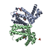
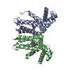
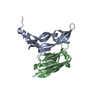
| Title | Crystal Structure Of The Yersinia enterocolitica Molecular Chaperone Syce | |||||||||
 Components Components | YOPE regulator | |||||||||
 Keywords Keywords | CHAPERONE / yersinia enterocolitica / molecular chaperone / type III secretion system | |||||||||
| Function / homology |  Function and homology information Function and homology informationprotein secretion by the type III secretion system / DNA-templated transcription Similarity search - Function | |||||||||
| Biological species |  Yersinia enterocolitica (bacteria) Yersinia enterocolitica (bacteria) | |||||||||
| Method |  X-RAY DIFFRACTION / SYNCHROTRON / MOLECULAR REPLACEMENT / Resolution: 2 Å X-RAY DIFFRACTION / SYNCHROTRON / MOLECULAR REPLACEMENT / Resolution: 2 Å | |||||||||
 Authors Authors | Trame, C.B. / McKay, D.B. | |||||||||
 Citation Citation | Journal: Acta Crystallogr.,Sect.D / Year: 2003 Title: Structure of the Yersinia enterocolitica molecular-chaperone protein SycE. Authors: Trame, C.B. / McKay, D.B. #1: Journal: NAT.STRUCT.BIOL. / Year: 2001Title: Structure of the Yersinia Type III Secretory System Chaperone Syce Authors: Birtalan, S. / Ghosh, P. #2: Journal: ACTA CRYSTALLOGR.,SECT.D / Year: 2002Title: Three-Dimensional Structure of the Type III Secretion Chaperone SycE from Yersinia Pestis Authors: Evdokimov, A.G. / Tropea, J.E. / Routzahn, K.M. / Waugh, D.S. #3: Journal: MOL.CELL / Year: 2002Title: Three-Dimensional Secretion Signals in Chaperone-Effector Complexes of Bacterial Pathogens Authors: Birtalan, S.C. / Phillips, R.M. / Ghosh, P. | |||||||||
| History |
|
- Structure visualization
| Structure viewer | Molecule: MolmilJmol/JSmol |
|---|
- Downloads & links
-Download
| PDBx/mmCIF format | 1n5b.cif.gz | 108.7 KB | Display | PDBx/mmCIF format |
|---|---|---|---|---|
| PDB format | pdb1n5b.ent.gz | 85.4 KB | Display | PDB format |
| PDBx/mmJSON format | 1n5b.json.gz | Tree view | PDBx/mmJSON format | |
| Others |  Other downloads Other downloads |
-Validation report
| Arichive directory | https://data.pdbj.org/pub/pdb/validation_reports/n5/1n5bftp://data.pdbj.org/pub/pdb/validation_reports/n5/1n5b | HTTPS FTP |
|---|
-Related structure data
| Related structure data |  1k6zS S: Starting model for refinement |
|---|---|
| Similar structure data |


-Links
 PDBj
PDBj
- Assembly
| Deposited unit | 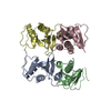
| ||||||||
|---|---|---|---|---|---|---|---|---|---|
| 1 | 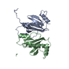
| ||||||||
| 2 | 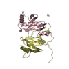
| ||||||||
| 3 | 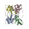
| ||||||||
| Unit cell |
| ||||||||
| Details | there are 4 molecules in AU, packed as 2 dimers with an ncs translation peak in the native patterson (0.5,~0.,0.5) |
-Components
| #1: Protein | Mass: 14839.753 Da / Num. of mol.: 4 Source method: isolated from a genetically manipulated source Source: (gene. exp.) Yersinia enterocolitica (bacteria) / Gene: YERA / Plasmid: pTYB / Species (production host): Escherichia coli / Production host: #2: Water | ChemComp-HOH / |  Mass: 18.015 Da / Num. of mol.: 89 / Source method: isolated from a natural source / Formula: H2O Mass: 18.015 Da / Num. of mol.: 89 / Source method: isolated from a natural source / Formula: H2O |
|---|
-Experimental details
-Experiment
| Experiment | Method: X-RAY DIFFRACTION / Number of used crystals: 1 |
|---|
- Sample preparation
| Crystal | Density Matthews: 2.17 Å3/Da / Density % sol: 42.98 % |
|---|---|
| Crystal grow | Temperature: 291 K / Method: vapor diffusion, hanging drop / pH: 7.7 Details: PEG 400, ammonium sulphate, sodium chloride, hepes, TCEP, pH 7.7, VAPOR DIFFUSION, HANGING DROP, temperature 291K |
-Data collection
| Diffraction | Mean temperature: 100 K |
|---|---|
| Diffraction source | Source: SYNCHROTRON / Site: SSRL  / Beamline: BL11-1 / Wavelength: 0.98 Å / Beamline: BL11-1 / Wavelength: 0.98 Å |
| Detector | Type: ADSC QUANTUM 315 / Detector: CCD / Date: May 27, 2002 Details: vertical focusing flat mirror, single crystal Si(111) bent monochromator (horizontal focussing) |
| Radiation | Monochromator: horizontally focusing bent Si(111) crystal / Protocol: SINGLE WAVELENGTH / Monochromatic (M) / Laue (L): M / Scattering type: x-ray |
| Radiation wavelength | Wavelength: 0.98 Å / Relative weight: 1 |
| Reflection | Resolution: 1.98→45.72 Å / Num. obs: 34752 / Observed criterion σ(F): 1 / Observed criterion σ(I): 1 / Redundancy: 4.4 % / Biso Wilson estimate: 21.7 Å2 / Rmerge(I) obs: 0.037 / Rsym value: 0.037 / Net I/σ(I): 30.6 |
| Reflection shell | Resolution: 1.98→2.01 Å / Redundancy: 4 % / Rmerge(I) obs: 0.235 / Mean I/σ(I) obs: 5 / Rsym value: 0.235 / % possible all: 89.2 |
- Processing
| Software |
| ||||||||||||||||||||||||||||||||||||||||||||||||||||||||||||
|---|---|---|---|---|---|---|---|---|---|---|---|---|---|---|---|---|---|---|---|---|---|---|---|---|---|---|---|---|---|---|---|---|---|---|---|---|---|---|---|---|---|---|---|---|---|---|---|---|---|---|---|---|---|---|---|---|---|---|---|---|---|
| Refinement | Method to determine structure: MOLECULAR REPLACEMENT Starting model: PDB ENTRY 1K6Z Resolution: 2→29.54 Å / Rfactor Rfree error: 0.005 / Data cutoff high absF: 1519681.15 / Data cutoff low absF: 0 / Isotropic thermal model: RESTRAINED / Cross valid method: THROUGHOUT / σ(F): 0 / Stereochemistry target values: Engh & Huber / Details: BULK SOLVENT MODEL USED
| ||||||||||||||||||||||||||||||||||||||||||||||||||||||||||||
| Solvent computation | Solvent model: FLAT MODEL / Bsol: 41.8009 Å2 / ksol: 0.357917 e/Å3 | ||||||||||||||||||||||||||||||||||||||||||||||||||||||||||||
| Displacement parameters | Biso mean: 48.4 Å2
| ||||||||||||||||||||||||||||||||||||||||||||||||||||||||||||
| Refine analyze |
| ||||||||||||||||||||||||||||||||||||||||||||||||||||||||||||
| Refinement step | Cycle: LAST / Resolution: 2→29.54 Å
| ||||||||||||||||||||||||||||||||||||||||||||||||||||||||||||
| Refine LS restraints |
| ||||||||||||||||||||||||||||||||||||||||||||||||||||||||||||
| LS refinement shell | Resolution: 2→2.13 Å / Rfactor Rfree error: 0.014 / Total num. of bins used: 6
| ||||||||||||||||||||||||||||||||||||||||||||||||||||||||||||
| Xplor file |
|
