 Movie
Movie Controller


+ Open data

- Basic information
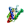
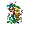
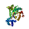
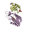
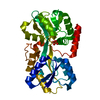
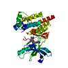
| Entry | Database: PDB / ID: 1k6z | ||||||
|---|---|---|---|---|---|---|---|
| Title | Crystal Structure of the Yersinia Secretion Chaperone SycE | ||||||
 Components Components | Type III secretion chaperone SycE | ||||||
 Keywords Keywords | CHAPERONE / secretion / yersinia pestis / toxin | ||||||
| Function / homology |  Function and homology information Function and homology informationprotein secretion by the type III secretion system / DNA-templated transcription Similarity search - Function | ||||||
| Biological species |   Yersinia pestis (bacteria) Yersinia pestis (bacteria) | ||||||
| Method |  X-RAY DIFFRACTION / SYNCHROTRON / MAD / Resolution: 2 Å X-RAY DIFFRACTION / SYNCHROTRON / MAD / Resolution: 2 Å | ||||||
 Authors Authors | Evdokimov, A.G. / Tropea, J.E. / Routzahn, K.M. / Waugh, D.S. | ||||||
 Citation Citation | Journal: Acta Crystallogr.,Sect.D / Year: 2002 Title: Three-dimensional structure of the type III secretion chaperone SycE from Yersinia pestis. Authors: Evdokimov, A.G. / Tropea, J.E. / Routzahn, K.M. / Waugh, D.S. | ||||||
| History |
|
- Structure visualization
| Structure viewer | Molecule: MolmilJmol/JSmol |
|---|
- Downloads & links
-Download
| PDBx/mmCIF format | 1k6z.cif.gz | 67.9 KB | Display | PDBx/mmCIF format |
|---|---|---|---|---|
| PDB format | pdb1k6z.ent.gz | 48.4 KB | Display | PDB format |
| PDBx/mmJSON format | 1k6z.json.gz | Tree view | PDBx/mmJSON format | |
| Others |  Other downloads Other downloads |
-Validation report
| Arichive directory | https://data.pdbj.org/pub/pdb/validation_reports/k6/1k6zftp://data.pdbj.org/pub/pdb/validation_reports/k6/1k6z | HTTPS FTP |
|---|
-Related structure data
| Related structure data | |
|---|---|
| Similar structure data |


-Links
 PDBj
PDBj


- Assembly
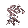
| Deposited unit | 
| ||||||||
|---|---|---|---|---|---|---|---|---|---|
| 1 |
| ||||||||
| Unit cell |
| ||||||||
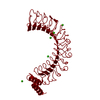
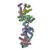
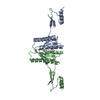
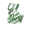
| Details | we believe that the biological assembly is a dimer, represented in the A.U. |
-Components
| #1: Protein | Mass: 16280.763 Da / Num. of mol.: 2 Source method: isolated from a genetically manipulated source Source: (gene. exp.) Yersinia pestis (bacteria) / Gene: SycE / Plasmid: pKM596 / Species (production host): Escherichia coli / Production host: #2: Chemical |   Mass: 69.085 Da / Num. of mol.: 2 / Source method: obtained synthetically / Formula: C3H5N2 Mass: 69.085 Da / Num. of mol.: 2 / Source method: obtained synthetically / Formula: C3H5N2#3: Water | ChemComp-HOH / |  Mass: 18.015 Da / Num. of mol.: 194 / Source method: isolated from a natural source / Formula: H2O Mass: 18.015 Da / Num. of mol.: 194 / Source method: isolated from a natural source / Formula: H2OHas protein modification | Y | |
|---|
-Experimental details
-Experiment
| Experiment | Method: X-RAY DIFFRACTION / Number of used crystals: 1 |
|---|
- Sample preparation
| Crystal | Density Matthews: 2.24 Å3/Da / Density % sol: 45.11 % | ||||||||||||||||||
|---|---|---|---|---|---|---|---|---|---|---|---|---|---|---|---|---|---|---|---|
| Crystal grow | Temperature: 300 K / Method: vapor diffusion, hanging drop / pH: 7.3 Details: 20% PEG8000, 500 mM Imidazole Acetate pH 7.3, VAPOR DIFFUSION, HANGING DROP, temperature 300K | ||||||||||||||||||
| Crystal grow | *PLUS Method: vapor diffusion | ||||||||||||||||||
| Components of the solutions | *PLUS
|
-Data collection
| Diffraction | Mean temperature: 100 K | ||||||||||||
|---|---|---|---|---|---|---|---|---|---|---|---|---|---|
| Diffraction source | Source: SYNCHROTRON / Site: NSLS  / Beamline: X9B / Wavelength: 0.9796, 0.9795, 0.9400 / Beamline: X9B / Wavelength: 0.9796, 0.9795, 0.9400 | ||||||||||||
| Detector | Type: ADSC QUANTUM 4 / Detector: CCD / Date: May 10, 2001 | ||||||||||||
| Radiation | Monochromator: X9B silicon / Protocol: MAD / Monochromatic (M) / Laue (L): M / Scattering type: x-ray | ||||||||||||
| Radiation wavelength |
| ||||||||||||
| Reflection | Resolution: 2→35 Å / Num. obs: 16201 / % possible obs: 82 % / Observed criterion σ(F): 0 / Observed criterion σ(I): 0 / Redundancy: 1.92 % / Biso Wilson estimate: 28 Å2 / Rmerge(I) obs: 0.04 / Rsym value: 0.045 / Net I/σ(I): 16.65 | ||||||||||||
| Reflection shell | Resolution: 2→2.05 Å / Redundancy: 1.7 % / Rmerge(I) obs: 0.27 / Mean I/σ(I) obs: 3.6 / Num. unique all: 1372 / % possible all: 99 | ||||||||||||
| Reflection | *PLUS Highest resolution: 1.95 Å / Lowest resolution: 30 Å / Num. obs: 21311 / % possible obs: 99.9 % / Redundancy: 1.9 % / Rmerge(I) obs: 0.05 | ||||||||||||
| Reflection shell | *PLUS % possible obs: 99.7 % / Rmerge(I) obs: 0.272 / Mean I/σ(I) obs: 2.3 |
- Processing
| Software |
| |||||||||||||||||||||||||
|---|---|---|---|---|---|---|---|---|---|---|---|---|---|---|---|---|---|---|---|---|---|---|---|---|---|---|
| Refinement | Method to determine structure: MAD Starting model: MAD Resolution: 2→35 Å / Isotropic thermal model: one per atom / Cross valid method: THROUGHOUT / σ(F): 0 / σ(I): 0 / Stereochemistry target values: Engh & Huber / Details: conjugated-gradient LS procedure in SHELXL
| |||||||||||||||||||||||||
| Displacement parameters | Biso mean: 29 Å2 | |||||||||||||||||||||||||
| Refinement step | Cycle: LAST / Resolution: 2→35 Å
| |||||||||||||||||||||||||
| Refine LS restraints |
| |||||||||||||||||||||||||
| Software | *PLUS Name: SHELXL-97 / Classification: refinement | |||||||||||||||||||||||||
| Refinement | *PLUS Highest resolution: 2 Å / Lowest resolution: 35 Å / σ(F): 0 / % reflection Rfree: 5 % / Rfactor obs: 0.194 | |||||||||||||||||||||||||
| Solvent computation | *PLUS | |||||||||||||||||||||||||
| Displacement parameters | *PLUS | |||||||||||||||||||||||||
| Refine LS restraints | *PLUS
|
