 Movie
Movie Controller

[English] 日本語
 Yorodumi
- PDB-1l2w: Crystal Structure of the Yersinia Virulence Effector YopE Chapero... -
+ Open data

- Basic information
| Entry | Database: PDB / ID: 1l2w | ||||||
|---|---|---|---|---|---|---|---|
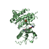
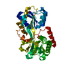
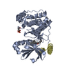
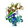
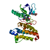
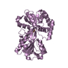
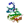
| Title | Crystal Structure of the Yersinia Virulence Effector YopE Chaperone-binding Domain in Complex with its Secretion Chaperone, SycE | ||||||
 Components Components |
| ||||||
 Keywords Keywords | CHAPERONE / CHAPERONE AND VIRULENCE PROTEIN | ||||||
| Function / homology |  Function and homology information Function and homology informationprotein secretion by the type III secretion system / negative regulation of phagocytosis / GTPase activator activity / cell outer membrane Similarity search - Function | ||||||
| Biological species |  Yersinia pseudotuberculosis (bacteria) Yersinia pseudotuberculosis (bacteria) | ||||||
| Method |  X-RAY DIFFRACTION / SYNCHROTRON / MOLECULAR REPLACEMENT / Resolution: 2 Å X-RAY DIFFRACTION / SYNCHROTRON / MOLECULAR REPLACEMENT / Resolution: 2 Å | ||||||
 Authors Authors | Birtalan, S.C. / Phillips, R.M. / Ghosh, P. | ||||||
 Citation Citation | Journal: Mol.Cell / Year: 2002 Title: Three-dimensional secretion signals in chaperone-effector complexes of bacterial pathogens. Authors: Birtalan, S.C. / Phillips, R.M. / Ghosh, P. | ||||||
| History |
| ||||||
| Remark 999 | SEQUENCE THE SEQUENCE OF THE SYCE PROTEIN, CHAINS A-H, MATCHES SWISS PROT ENTRY P31491, WHOSE ...SEQUENCE THE SEQUENCE OF THE SYCE PROTEIN, CHAINS A-H, MATCHES SWISS PROT ENTRY P31491, WHOSE SOURCE IS YERSINIA PESTIS. THE SOURCE OF THE SYCE PROTEIN IN THIS ENTRY IS YERSINIA PSEUDOTUBERCULOSIS. THERE IS AN EXTRA GLYCINE, RESIDUE 1, IN CHAINS A-H WHICH WAS INSERTED FOR CLONING PURPOSES, and the last 8 residues were cleaved to yield residues 0-122. The N- and C- terminal residues of chains I-L were cleaved to yield residues 17-85. |
- Structure visualization
| Structure viewer | Molecule: MolmilJmol/JSmol |
|---|
- Downloads & links
-Download
| PDBx/mmCIF format | 1l2w.cif.gz | 240.6 KB | Display | PDBx/mmCIF format |
|---|---|---|---|---|
| PDB format | pdb1l2w.ent.gz | 196.2 KB | Display | PDB format |
| PDBx/mmJSON format | 1l2w.json.gz | Tree view | PDBx/mmJSON format | |
| Others |  Other downloads Other downloads |
-Validation report
| Arichive directory | https://data.pdbj.org/pub/pdb/validation_reports/l2/1l2wftp://data.pdbj.org/pub/pdb/validation_reports/l2/1l2w | HTTPS FTP |
|---|
-Related structure data
| Related structure data | 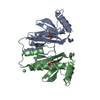 1jyaS S: Starting model for refinement |
|---|---|
| Similar structure data |

-Links
 PDBj
PDBj
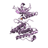
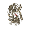
- Assembly
| Deposited unit | 
| ||||||||
|---|---|---|---|---|---|---|---|---|---|
| 1 | 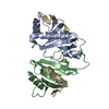
| ||||||||
| 2 | 
| ||||||||
| 3 | 
| ||||||||
| 4 | 
| ||||||||
| 5 | 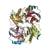
| ||||||||
| 6 | 
| ||||||||
| Unit cell |
|
-Components
| #1: Protein | Mass: 13843.607 Da / Num. of mol.: 8 Source method: isolated from a genetically manipulated source Source: (gene. exp.) Yersinia pseudotuberculosis (bacteria) / Gene: syce / Plasmid: pET28b / Species (production host): Escherichia coli / Production host: #2: Protein | Mass: 7277.952 Da / Num. of mol.: 4 / Fragment: Chaperone-binding Domain Source method: isolated from a genetically manipulated source Source: (gene. exp.) Yersinia pseudotuberculosis (bacteria) / Gene: yope / Plasmid: pET28b / Species (production host): Escherichia coli / Production host: #3: Water | ChemComp-HOH / |  Mass: 18.015 Da / Num. of mol.: 362 / Source method: isolated from a natural source / Formula: H2O Mass: 18.015 Da / Num. of mol.: 362 / Source method: isolated from a natural source / Formula: H2O |
|---|
-Experimental details
-Experiment
| Experiment | Method: X-RAY DIFFRACTION / Number of used crystals: 1 |
|---|
- Sample preparation
| Crystal | Density Matthews: 2.38 Å3/Da / Density % sol: 48.32 % | ||||||||||||||||||||||||||||||||||||||||||||||||
|---|---|---|---|---|---|---|---|---|---|---|---|---|---|---|---|---|---|---|---|---|---|---|---|---|---|---|---|---|---|---|---|---|---|---|---|---|---|---|---|---|---|---|---|---|---|---|---|---|---|
| Crystal grow | Temperature: 291 K / Method: vapor diffusion, hanging drop / pH: 4.6 Details: PEG 8000, sodium tartrate, sodium acetate, dithiothreitol, pH 4.6, VAPOR DIFFUSION, HANGING DROP, temperature 291K | ||||||||||||||||||||||||||||||||||||||||||||||||
| Crystal | *PLUS Density % sol: 50 % | ||||||||||||||||||||||||||||||||||||||||||||||||
| Crystal grow | *PLUS Temperature: 18 ℃ / pH: 8 / Method: unknown | ||||||||||||||||||||||||||||||||||||||||||||||||
| Components of the solutions | *PLUS
|
-Data collection
| Diffraction | Mean temperature: 100 K |
|---|---|
| Diffraction source | Source: SYNCHROTRON / Site: ALS  / Beamline: 5.0.2 / Wavelength: 1 Å / Beamline: 5.0.2 / Wavelength: 1 Å |
| Detector | Type: ADSC QUANTUM 4 / Detector: CCD / Date: Mar 30, 2001 |
| Radiation | Protocol: SINGLE WAVELENGTH / Monochromatic (M) / Laue (L): M / Scattering type: x-ray |
| Radiation wavelength | Wavelength: 1 Å / Relative weight: 1 |
| Reflection | Resolution: 1.99→20 Å / Num. all: 87145 / Num. obs: 84804 / % possible obs: 97.3 % / Observed criterion σ(F): 0 / Observed criterion σ(I): -3 / Redundancy: 2 % / Biso Wilson estimate: 36.69 Å2 / Rmerge(I) obs: 0.058 / Rsym value: 0.058 / Net I/σ(I): 18.22 |
| Reflection shell | Resolution: 1.99→2.03 Å / Redundancy: 2 % / Rmerge(I) obs: 0.46 / Mean I/σ(I) obs: 1.77 / Num. unique all: 4425 / Rsym value: 0.46 / % possible all: 96.5 |
| Reflection | *PLUS Highest resolution: 2.03 Å / Lowest resolution: 20 Å / % possible obs: 96.7 % |
| Reflection shell | *PLUS Highest resolution: 2.03 Å / Lowest resolution: 2.07 Å / % possible obs: 96.7 % / Rmerge(I) obs: 0.49 / Mean I/σ(I) obs: 2.1 |
- Processing
| Software |
| |||||||||||||||||||||||||
|---|---|---|---|---|---|---|---|---|---|---|---|---|---|---|---|---|---|---|---|---|---|---|---|---|---|---|
| Refinement | Method to determine structure: MOLECULAR REPLACEMENT Starting model: PDB entry 1jya Resolution: 2→20 Å / Isotropic thermal model: atomic / Cross valid method: THROUGHOUT / σ(F): 0 / σ(I): 0 / Stereochemistry target values: Engh & Huber
| |||||||||||||||||||||||||
| Displacement parameters | Biso mean: 48.05 Å2
| |||||||||||||||||||||||||
| Refine analyze |
| |||||||||||||||||||||||||
| Refinement step | Cycle: LAST / Resolution: 2→20 Å
| |||||||||||||||||||||||||
| Refine LS restraints |
| |||||||||||||||||||||||||
| LS refinement shell | Resolution: 2→2.07 Å
| |||||||||||||||||||||||||
| Refinement | *PLUS Highest resolution: 2.03 Å / Lowest resolution: 20 Å / Num. reflection obs: 77053 / Num. reflection Rfree: 4091 / % reflection Rfree: 5 % / Rfactor obs: 0.249 / Rfactor Rfree: 0.274 / Rfactor Rwork: 0.249 | |||||||||||||||||||||||||
| Solvent computation | *PLUS | |||||||||||||||||||||||||
| Displacement parameters | *PLUS | |||||||||||||||||||||||||
| Refine LS restraints | *PLUS
| |||||||||||||||||||||||||
| LS refinement shell | *PLUS Rfactor obs: 0.3497 |
