 Movie
Movie Controller


+ Open data

- Basic information
| Entry | Database: PDB / ID: 1n1a | ||||||
|---|---|---|---|---|---|---|---|
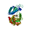
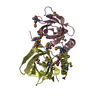
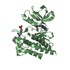
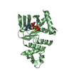
| Title | Crystal Structure of the N-terminal domain of human FKBP52 | ||||||
 Components Components | FKBP52 | ||||||
 Keywords Keywords | ISOMERASE / FKBP52 / the N-terminal domain | ||||||
| Function / homology |  Function and homology information Function and homology informationsteroid hormone receptor complex assembly / negative regulation of microtubule polymerization or depolymerization / male sex differentiation / copper-dependent protein binding / prostate gland development / copper ion transport / nuclear glucocorticoid receptor binding / protein-containing complex localization / androgen receptor signaling pathway / FK506 binding ...steroid hormone receptor complex assembly / negative regulation of microtubule polymerization or depolymerization / male sex differentiation / copper-dependent protein binding / prostate gland development / copper ion transport / nuclear glucocorticoid receptor binding / protein-containing complex localization / androgen receptor signaling pathway / FK506 binding / negative regulation of microtubule polymerization / Attenuation phase / axonal growth cone / heat shock protein binding / embryo implantation / ESR-mediated signaling / HSP90 chaperone cycle for steroid hormone receptors (SHR) in the presence of ligand / peptidylprolyl isomerase / peptidyl-prolyl cis-trans isomerase activity / phosphoprotein binding / tau protein binding / negative regulation of neuron projection development / protein folding / Estrogen-dependent gene expression / Potential therapeutics for SARS / microtubule / protein-macromolecule adaptor activity / neuronal cell body / GTP binding / perinuclear region of cytoplasm / protein-containing complex / mitochondrion / RNA binding / extracellular exosome / nucleoplasm / ATP binding / cytoplasm / cytosol Similarity search - Function | ||||||
| Biological species |  Homo sapiens (human) Homo sapiens (human) | ||||||
| Method |  X-RAY DIFFRACTION / MOLECULAR REPLACEMENT / Resolution: 2.4 Å X-RAY DIFFRACTION / MOLECULAR REPLACEMENT / Resolution: 2.4 Å | ||||||
 Authors Authors | Li, P. / Ding, Y. / Wu, B. / Shu, C. / Shen, B. / Rao, Z. | ||||||
 Citation Citation | Journal: Acta Crystallogr.,Sect.D / Year: 2003 Title: Structure of the N-terminal domain of human FKBP52. Authors: Li, P. / Ding, Y. / Wu, B. / Shu, C. / Shen, B. / Rao, Z. | ||||||
| History |
|
- Structure visualization
| Structure viewer | Molecule: MolmilJmol/JSmol |
|---|
- Downloads & links
-Download
| PDBx/mmCIF format | 1n1a.cif.gz | 58.7 KB | Display | PDBx/mmCIF format |
|---|---|---|---|---|
| PDB format | pdb1n1a.ent.gz | 43 KB | Display | PDB format |
| PDBx/mmJSON format | 1n1a.json.gz | Tree view | PDBx/mmJSON format | |
| Others |  Other downloads Other downloads |
-Validation report
| Arichive directory | https://data.pdbj.org/pub/pdb/validation_reports/n1/1n1aftp://data.pdbj.org/pub/pdb/validation_reports/n1/1n1a | HTTPS FTP |
|---|
-Related structure data
| Related structure data | 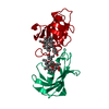 1fkjS S: Starting model for refinement |
|---|---|
| Similar structure data |


-Links
 PDBj
PDBj


- Assembly
| Deposited unit | 
| ||||||||
|---|---|---|---|---|---|---|---|---|---|
| 1 |
| ||||||||
| Unit cell |
| ||||||||
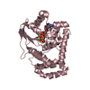
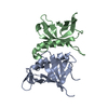
| Details | The biological assembly is a dimer in the asymmetric unit |
-Components
| #1: Protein | Mass: 15336.519 Da / Num. of mol.: 2 / Fragment: the N-terminal fragment (1-140) Source method: isolated from a genetically manipulated source Source: (gene. exp.) Homo sapiens (human) / Gene: FKBP4 / Plasmid: PET28a(+) / Species (production host): Escherichia coli / Production host:  #2: Water | ChemComp-HOH / |  Mass: 18.015 Da / Num. of mol.: 55 / Source method: isolated from a natural source / Formula: H2O Mass: 18.015 Da / Num. of mol.: 55 / Source method: isolated from a natural source / Formula: H2O |
|---|
-Experimental details
-Experiment
| Experiment | Method: X-RAY DIFFRACTION / Number of used crystals: 1 |
|---|
- Sample preparation
| Crystal | Density Matthews: 1.85 Å3/Da / Density % sol: 33.68 % | ||||||||||||||||||||||||||||||
|---|---|---|---|---|---|---|---|---|---|---|---|---|---|---|---|---|---|---|---|---|---|---|---|---|---|---|---|---|---|---|---|
| Crystal grow | Temperature: 291 K / Method: vapor diffusion, hanging drop / pH: 8.5 Details: Tris-HCl, Ammonium Sulfate, pH 8.5, VAPOR DIFFUSION, HANGING DROP, temperature 291K | ||||||||||||||||||||||||||||||
| Crystal grow | *PLUS | ||||||||||||||||||||||||||||||
| Components of the solutions | *PLUS
|
-Data collection
| Diffraction | Mean temperature: 100 K |
|---|---|
| Diffraction source | Source: ROTATING ANODE / Type: RIGAKU / Wavelength: 1.5418 Å |
| Detector | Type: MARRESEARCH / Detector: IMAGE PLATE / Date: Jan 1, 2001 / Details: mirrors |
| Radiation | Monochromator: CuKa / Protocol: SINGLE WAVELENGTH / Monochromatic (M) / Laue (L): M / Scattering type: x-ray |
| Radiation wavelength | Wavelength: 1.5418 Å / Relative weight: 1 |
| Reflection | Resolution: 2.4→40 Å / Num. all: 26262 / Num. obs: 26259 / % possible obs: 99.9 % / Observed criterion σ(F): 1 / Observed criterion σ(I): 1 / Redundancy: 10.8 % / Rmerge(I) obs: 0.095 / Net I/σ(I): 17.3 |
| Reflection shell | Resolution: 2.4→2.49 Å / Redundancy: 8.4 % / Rmerge(I) obs: 0.294 / Mean I/σ(I) obs: 9.3 / Num. unique all: 8929 / % possible all: 99.9 |
| Reflection | *PLUS Lowest resolution: 40 Å / Num. obs: 8929 / Num. measured all: 26262 |
| Reflection shell | *PLUS Highest resolution: 2.4 Å / % possible obs: 99.9 % / Num. unique obs: 889 |
- Processing
| Software |
| |||||||||||||||||||||||||
|---|---|---|---|---|---|---|---|---|---|---|---|---|---|---|---|---|---|---|---|---|---|---|---|---|---|---|
| Refinement | Method to determine structure: MOLECULAR REPLACEMENT Starting model: PDB ENTRY 1FKJ Resolution: 2.4→40 Å / σ(F): 0 / σ(I): 0 / Stereochemistry target values: Engh & Huber
| |||||||||||||||||||||||||
| Displacement parameters |
| |||||||||||||||||||||||||
| Refinement step | Cycle: LAST / Resolution: 2.4→40 Å
| |||||||||||||||||||||||||
| Refine LS restraints |
| |||||||||||||||||||||||||
| Xplor file |
| |||||||||||||||||||||||||
| Refinement | *PLUS Lowest resolution: 40 Å | |||||||||||||||||||||||||
| Solvent computation | *PLUS | |||||||||||||||||||||||||
| Displacement parameters | *PLUS | |||||||||||||||||||||||||
| Refine LS restraints | *PLUS
|
