 Movie
Movie Controller

[English] 日本語
 Yorodumi
- PDB-1n14: Structure and Dynamics of Thioguanine-modified Duplex DNA in Comp... -
+ Open data

- Basic information
| Entry | Database: PDB / ID: 1n14 | ||||||
|---|---|---|---|---|---|---|---|
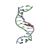
| Title | Structure and Dynamics of Thioguanine-modified Duplex DNA in Comparison with Unmodified DNA; Structure of Unmodified Duplex DNA | ||||||
 Components Components |
| ||||||
 Keywords Keywords | DNA / mercaptopurine / thioguanine / anti-cancer therapy | ||||||
| Function / homology | DNA / DNA (> 10) Function and homology information Function and homology information | ||||||
| Method | SOLUTION NMR / restrained molecular dynamics, relaxation matrix analysis | ||||||
 Authors Authors | Somerville, L. / Krynetski, E.Y. / Krynetskaia, N.F. / Beger, R.D. / Zhang, W. / Marhefka, C.A. / Evans, W.E. / Kriwacki, R.W. | ||||||
 Citation Citation | Journal: J.Biol.Chem. / Year: 2003 Title: Structure and dynamics of thioguanine-modified duplex DNA Authors: Somerville, L. / Krynetski, E.Y. / Krynetskaia, N.F. / Beger, R.D. / Zhang, W. / Marhefka, C.A. / Evans, W.E. / Kriwacki, R.W. | ||||||
| History |
|
- Structure visualization
| Structure viewer | Molecule: MolmilJmol/JSmol |
|---|
- Downloads & links
-Download
| PDBx/mmCIF format | 1n14.cif.gz | 379.6 KB | Display | PDBx/mmCIF format |
|---|---|---|---|---|
| PDB format | pdb1n14.ent.gz | 272 KB | Display | PDB format |
| PDBx/mmJSON format | 1n14.json.gz | Tree view | PDBx/mmJSON format | |
| Others |  Other downloads Other downloads |
-Validation report
| Arichive directory | https://data.pdbj.org/pub/pdb/validation_reports/n1/1n14ftp://data.pdbj.org/pub/pdb/validation_reports/n1/1n14 | HTTPS FTP |
|---|
-Related structure data


-Links
 PDBj
PDBj


- Assembly
| Deposited unit | 
| |||||||||
|---|---|---|---|---|---|---|---|---|---|---|
| 1 |
| |||||||||
| NMR ensembles |
|
-Components
| #1: DNA chain | Mass: 4009.637 Da / Num. of mol.: 1 / Source method: obtained synthetically |
|---|---|
| #2: DNA chain | Mass: 3933.558 Da / Num. of mol.: 1 / Source method: obtained synthetically |
-Experimental details
-Experiment
| Experiment | Method: SOLUTION NMR | ||||||||||||||||
|---|---|---|---|---|---|---|---|---|---|---|---|---|---|---|---|---|---|
| NMR experiment |
|
- Sample preparation
| Details | Contents: Single-stranded oligodeoxyribonucleotides, d(5-GCTAAGGAAAGCC-3) and the complementary strand d(5-GGCTTTCCTTAGC-3), were synthesized using standard phosphoramidite chemistry. Single-stranded ...Contents: Single-stranded oligodeoxyribonucleotides, d(5-GCTAAGGAAAGCC-3) and the complementary strand d(5-GGCTTTCCTTAGC-3), were synthesized using standard phosphoramidite chemistry. Single-stranded DNA molecules were purified using anion exchange chromatography (MonoQ HR 5/5, Pharmacia Biotech). Purity was confirmed by UV spectroscopy, analytical anion exchange chromatography, and mass spectrometry. The GC and thioGC DNA duplexes were each prepared by annealing either equimolar amounts of complimentary strands or a 1.2 molar excess of the unmodified strand to the modified strand in Buffer A (10 mM sodium phosphate, 50 mM NaCl, pH 7.0) at 70 C for 5 min., followed by slow cooling over 12 hours to room temperature. DNA duplexes were purified using gel filtration chromatography (Superdex Peptide HR 10/30, Pharmacia Biotech) in Buffer A. Solvent system: Buffer A (10 mM sodium phosphate, 50 mM NaCl, pH 7.0) |
|---|---|
| Sample conditions | Ionic strength: approx. 65 mM / pH: 7 / Pressure: ambient / Temperature: 293 K |
| Crystal grow | *PLUS Method: other / Details: NMR |
-NMR measurement
| Radiation | Protocol: SINGLE WAVELENGTH / Monochromatic (M) / Laue (L): M | |||||||||||||||
|---|---|---|---|---|---|---|---|---|---|---|---|---|---|---|---|---|
| Radiation wavelength | Relative weight: 1 | |||||||||||||||
| NMR spectrometer |
|
- Processing
| NMR software |
| ||||||||||||
|---|---|---|---|---|---|---|---|---|---|---|---|---|---|
| Refinement | Method: restrained molecular dynamics, relaxation matrix analysis Software ordinal: 1 Details: Restrained molecular dynamics (rMD) structure refinement of B-DNA starting structures was performed using an XPLOR 3.1 (26) simulated annealing protocol employing the Cheatham, et al. (19), ...Details: Restrained molecular dynamics (rMD) structure refinement of B-DNA starting structures was performed using an XPLOR 3.1 (26) simulated annealing protocol employing the Cheatham, et al. (19), force field. This force field was modified to include parameterization of thioG, as described above. Starting structures were energy minimized by 160 steps of Powells conjugate gradient minimization followed by rMD while heating to 600 K at 50 K sec-1, cooling to 300 K at 25 K sec-1, and equilibrating at 293 K over 250 ps. | ||||||||||||
| NMR ensemble | Conformer selection criteria: back calculated data agree with experimental NOESY spectrum Conformers calculated total number: 200 / Conformers submitted total number: 20 |
 X-PLOR