 Movie
Movie Controller

[English] 日本語
 Yorodumi
- PDB-1z1x: Crystal Structure of a novel disintegrin from Saw-scaled viper at... -
+ Open data

- Basic information
| Entry | Database: PDB / ID: 1z1x | ||||||
|---|---|---|---|---|---|---|---|
| Title | Crystal Structure of a novel disintegrin from Saw-scaled viper at 3.2 A resolution | ||||||
 Components Components | disintegrin | ||||||
 Keywords Keywords | PROTEIN BINDING / DISINTEGRIN / SAW-SCALED VIPER | ||||||
| Function / homology |  Function and homology information Function and homology information | ||||||
| Biological species |  Echis carinatus (saw-scaled viper) Echis carinatus (saw-scaled viper) | ||||||
| Method |  X-RAY DIFFRACTION / MOLECULAR REPLACEMENT / Resolution: 3.2 Å X-RAY DIFFRACTION / MOLECULAR REPLACEMENT / Resolution: 3.2 Å | ||||||
 Authors Authors | Hassan, M.I. / Ethayathulla, A.S. / Bilgrami, S. / Singh, B. / Yadav, S. / Singh, T.P. | ||||||
 Citation Citation | Journal: To be published Title: Crystal Structure of a novel disintegrin from Saw-scaled viper at 3.2A resolution Authors: Hassan, M.I. / Ethayathulla, A.S. / Bilgrami, S. / Singh, B. / Yadav, S. / Singh, T.P. | ||||||
| History |
|
- Structure visualization
| Structure viewer | Molecule: MolmilJmol/JSmol |
|---|
- Downloads & links
-Download
| PDBx/mmCIF format | 1z1x.cif.gz | 23.7 KB | Display | PDBx/mmCIF format |
|---|---|---|---|---|
| PDB format | pdb1z1x.ent.gz | 15.4 KB | Display | PDB format |
| PDBx/mmJSON format | 1z1x.json.gz | Tree view | PDBx/mmJSON format | |
| Others |  Other downloads Other downloads |
-Validation report
| Arichive directory | https://data.pdbj.org/pub/pdb/validation_reports/z1/1z1xftp://data.pdbj.org/pub/pdb/validation_reports/z1/1z1x | HTTPS FTP |
|---|
-Related structure data
| Related structure data |  1rmrS S: Starting model for refinement |
|---|---|
| Similar structure data |


-Links
 PDBj
PDBj- Assembly
| Deposited unit | 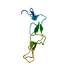
| ||||||||
|---|---|---|---|---|---|---|---|---|---|
| 1 | 
| ||||||||
| Unit cell |
|
-Components
| #1: Protein | Mass: 7139.176 Da / Num. of mol.: 1 / Source method: isolated from a natural source / Source: (natural) Echis carinatus (saw-scaled viper) / Secretion: Venom / Strain: Saw-scaled Viper / References: UniProt: Q5EE07 |
|---|---|
| #2: Water | ChemComp-HOH /  Mass: 18.015 Da / Num. of mol.: 8 / Source method: isolated from a natural source / Formula: H2O Mass: 18.015 Da / Num. of mol.: 8 / Source method: isolated from a natural source / Formula: H2O |
| Has protein modification | Y |
-Experimental details
-Experiment
| Experiment | Method: X-RAY DIFFRACTION / Number of used crystals: 1 |
|---|
- Sample preparation
| Crystal | Density Matthews: 4 Å3/Da / Density % sol: 70 % |
|---|---|
| Crystal grow | Temperature: 298 K / Method: vapor diffusion, sitting drop / pH: 6 Details: 1.2M AMMONIUM SULFATE, pH 6.0, VAPOR DIFFUSION, SITTING DROP, temperature 298K |
-Data collection
| Diffraction | Mean temperature: 298 K |
|---|---|
| Diffraction source | Source: ROTATING ANODE / Type: RIGAKU RU300 / Wavelength: 1.5418 Å |
| Detector | Type: MARRESEARCH / Detector: IMAGE PLATE / Date: Feb 10, 2005 / Details: MIRROR |
| Radiation | Monochromator: GRAPHITE / Protocol: SINGLE WAVELENGTH / Monochromatic (M) / Laue (L): M / Scattering type: x-ray |
| Radiation wavelength | Wavelength: 1.5418 Å / Relative weight: 1 |
| Reflection | Resolution: 3.2→64.55 Å / Num. all: 2062 / Num. obs: 2058 / % possible obs: 99.6 % / Observed criterion σ(F): 0 / Observed criterion σ(I): 0 / Redundancy: 10 % / Biso Wilson estimate: 55.2 Å2 / Rsym value: 0.2 / Net I/σ(I): 8.3 |
| Reflection shell | Resolution: 3.2→3.32 Å / Mean I/σ(I) obs: 3.9 / Num. unique all: 191 / Rsym value: 0.544 / % possible all: 97.6 |
- Processing
| Software |
| ||||||||||||||||||||||||||||||||||||||||||||||||||||||||||||||||||||||||||||||||||||||||||||||||||||
|---|---|---|---|---|---|---|---|---|---|---|---|---|---|---|---|---|---|---|---|---|---|---|---|---|---|---|---|---|---|---|---|---|---|---|---|---|---|---|---|---|---|---|---|---|---|---|---|---|---|---|---|---|---|---|---|---|---|---|---|---|---|---|---|---|---|---|---|---|---|---|---|---|---|---|---|---|---|---|---|---|---|---|---|---|---|---|---|---|---|---|---|---|---|---|---|---|---|---|---|---|---|
| Refinement | Method to determine structure: MOLECULAR REPLACEMENT Starting model: 1rmr Resolution: 3.2→64 Å / Cor.coef. Fo:Fc: 0.873 / Cor.coef. Fo:Fc free: 0.821 / SU B: 27.591 / SU ML: 0.486 / Cross valid method: THROUGHOUT / ESU R Free: 0.442 / Stereochemistry target values: MAXIMUM LIKELIHOOD / Details: HYDROGENS HAVE BEEN ADDED IN THE RIDING POSITIONS
| ||||||||||||||||||||||||||||||||||||||||||||||||||||||||||||||||||||||||||||||||||||||||||||||||||||
| Solvent computation | Ion probe radii: 0.8 Å / Shrinkage radii: 0.8 Å / VDW probe radii: 1.4 Å / Solvent model: BABINET MODEL WITH MASK | ||||||||||||||||||||||||||||||||||||||||||||||||||||||||||||||||||||||||||||||||||||||||||||||||||||
| Displacement parameters | Biso mean: 50.852 Å2
| ||||||||||||||||||||||||||||||||||||||||||||||||||||||||||||||||||||||||||||||||||||||||||||||||||||
| Refinement step | Cycle: LAST / Resolution: 3.2→64 Å
| ||||||||||||||||||||||||||||||||||||||||||||||||||||||||||||||||||||||||||||||||||||||||||||||||||||
| Refine LS restraints |
| ||||||||||||||||||||||||||||||||||||||||||||||||||||||||||||||||||||||||||||||||||||||||||||||||||||
| LS refinement shell | Resolution: 3.202→3.285 Å / Total num. of bins used: 20
|
