 Movie
Movie Controller

[English] 日本語
 Yorodumi
- PDB-1n12: Crystal structure of the PapE (N-terminal-deleted) pilus subunit ... -
+ Open data

- Basic information
| Entry | Database: PDB / ID: 1n12 | ||||||
|---|---|---|---|---|---|---|---|
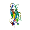
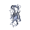
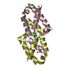
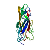
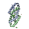
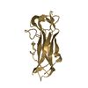
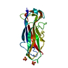
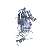
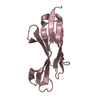
| Title | Crystal structure of the PapE (N-terminal-deleted) pilus subunit bound to a peptide corresponding to the N-terminal extension of the PapK pilus subunit (residues 1-11) from uropathogenic E. coli | ||||||
 Components Components |
| ||||||
 Keywords Keywords | CHAPERONE / Immunoglobulin-like fold / donor strand complementation / donor strand exchange / chaperone priming / pilus fiber assembly / organelle biogenesis | ||||||
| Function / homology |  Function and homology information Function and homology informationcell adhesion involved in single-species biofilm formation / pilus / cell adhesion / extracellular region Similarity search - Function | ||||||
| Biological species |  | ||||||
| Method |  X-RAY DIFFRACTION / SYNCHROTRON / SAD / Resolution: 1.87 Å X-RAY DIFFRACTION / SYNCHROTRON / SAD / Resolution: 1.87 Å | ||||||
 Authors Authors | Sauer, F.G. / Pinkner, J.S. / Waksman, G. / Hultgren, S.J. | ||||||
 Citation Citation | Journal: Cell(Cambridge,Mass.) / Year: 2002 Title: Chaperone priming of pilus subunits facilitates a topological transition that drives fiber formation Authors: Sauer, F.G. / Pinkner, J.S. / Waksman, G. / Hultgren, S.J. | ||||||
| History |
|
- Structure visualization
| Structure viewer | Molecule: MolmilJmol/JSmol |
|---|
- Downloads & links
-Download
| PDBx/mmCIF format | 1n12.cif.gz | 71.5 KB | Display | PDBx/mmCIF format |
|---|---|---|---|---|
| PDB format | pdb1n12.ent.gz | 53.1 KB | Display | PDB format |
| PDBx/mmJSON format | 1n12.json.gz | Tree view | PDBx/mmJSON format | |
| Others |  Other downloads Other downloads |
-Validation report
| Arichive directory | https://data.pdbj.org/pub/pdb/validation_reports/n1/1n12ftp://data.pdbj.org/pub/pdb/validation_reports/n1/1n12 | HTTPS FTP |
|---|
-Related structure data


-Links
 PDBj
PDBj
- Assembly
| Deposited unit | 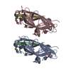
| ||||||||
|---|---|---|---|---|---|---|---|---|---|
| 1 | 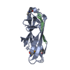
| ||||||||
| 2 | 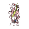
| ||||||||
| Unit cell |
|
-Components
| #1: Protein | Mass: 14832.035 Da / Num. of mol.: 2 / Fragment: residue 25-173, residue 26-36 deleted Mutation: N-terminal residue 26-36 deleted, contains selenomethionine at methionine positions Source method: isolated from a genetically manipulated source Source: (gene. exp.) #2: Protein/peptide | Mass: 1207.315 Da / Num. of mol.: 2 / Fragment: residue 22-32 / Source method: obtained synthetically / Details: synthesized peptide / References: UniProt: P42190, UniProt: P42191*PLUS #3: Water | ChemComp-HOH / |  Mass: 18.015 Da / Num. of mol.: 140 / Source method: isolated from a natural source / Formula: H2O Mass: 18.015 Da / Num. of mol.: 140 / Source method: isolated from a natural source / Formula: H2OHas protein modification | Y | |
|---|
-Experimental details
-Experiment
| Experiment | Method: X-RAY DIFFRACTION / Number of used crystals: 1 |
|---|
- Sample preparation
| Crystal | Density Matthews: 1.75 Å3/Da / Density % sol: 29.19 % |
|---|---|
| Crystal grow | Temperature: 294 K / Method: vapor diffusion / pH: 8.5 Details: 100 mM Tris pH 8.5, 200 mM NaCl, 27-32% PEG 4000, 1 mM beta-mercaptoethanol, VAPOR DIFFUSION, temperature 294K |
-Data collection
| Diffraction | Mean temperature: 98 K |
|---|---|
| Diffraction source | Source: SYNCHROTRON / Site: APS  / Beamline: 19-ID / Wavelength: 0.9667 / Beamline: 19-ID / Wavelength: 0.9667 |
| Radiation | Protocol: SINGLE WAVELENGTH / Monochromatic (M) / Laue (L): M / Scattering type: x-ray |
| Radiation wavelength | Wavelength: 0.9667 Å / Relative weight: 1 |
| Reflection | Biso Wilson estimate: 8 Å2 |
| Reflection | *PLUS Highest resolution: 1.87 Å / Lowest resolution: 30 Å / Num. obs: 18969 / % possible obs: 99.2 % / Num. measured all: 232749 / Rmerge(I) obs: 0.05 |
| Reflection shell | *PLUS % possible obs: 96.8 % / Rmerge(I) obs: 0.149 / Mean I/σ(I) obs: 8.4 |
- Processing
| Software |
| ||||||||||||||||||||||||||||||||||||||||||||||||||||||||||||||||||||||||||||||||
|---|---|---|---|---|---|---|---|---|---|---|---|---|---|---|---|---|---|---|---|---|---|---|---|---|---|---|---|---|---|---|---|---|---|---|---|---|---|---|---|---|---|---|---|---|---|---|---|---|---|---|---|---|---|---|---|---|---|---|---|---|---|---|---|---|---|---|---|---|---|---|---|---|---|---|---|---|---|---|---|---|---|
| Refinement | Method to determine structure: SAD / Resolution: 1.87→26.21 Å / Rfactor Rfree error: 0.007 / Isotropic thermal model: RESTRAINED / Cross valid method: THROUGHOUT / σ(F): 0
| ||||||||||||||||||||||||||||||||||||||||||||||||||||||||||||||||||||||||||||||||
| Solvent computation | Solvent model: FLAT MODEL / Bsol: 32.9399 Å2 / ksol: 0.348785 e/Å3 | ||||||||||||||||||||||||||||||||||||||||||||||||||||||||||||||||||||||||||||||||
| Displacement parameters | Biso mean: 19.9 Å2
| ||||||||||||||||||||||||||||||||||||||||||||||||||||||||||||||||||||||||||||||||
| Refine analyze | Luzzati coordinate error free: 0.28 Å / Luzzati sigma a free: 0.21 Å | ||||||||||||||||||||||||||||||||||||||||||||||||||||||||||||||||||||||||||||||||
| Refinement step | Cycle: LAST / Resolution: 1.87→26.21 Å
| ||||||||||||||||||||||||||||||||||||||||||||||||||||||||||||||||||||||||||||||||
| Refine LS restraints |
| ||||||||||||||||||||||||||||||||||||||||||||||||||||||||||||||||||||||||||||||||
| LS refinement shell | Resolution: 1.87→1.99 Å / Rfactor Rfree error: 0.021 / Total num. of bins used: 6
| ||||||||||||||||||||||||||||||||||||||||||||||||||||||||||||||||||||||||||||||||
| Xplor file |
| ||||||||||||||||||||||||||||||||||||||||||||||||||||||||||||||||||||||||||||||||
| Refinement | *PLUS Lowest resolution: 30 Å | ||||||||||||||||||||||||||||||||||||||||||||||||||||||||||||||||||||||||||||||||
| Solvent computation | *PLUS | ||||||||||||||||||||||||||||||||||||||||||||||||||||||||||||||||||||||||||||||||
| Displacement parameters | *PLUS | ||||||||||||||||||||||||||||||||||||||||||||||||||||||||||||||||||||||||||||||||
| Refine LS restraints | *PLUS
|
