 Movie
Movie Controller

[English] 日本語
 Yorodumi
- PDB-1mzw: Crystal structure of a U4/U6 snRNP complex between human spliceos... -
+ Open data

- Basic information
| Entry | Database: PDB / ID: 1mzw | ||||||
|---|---|---|---|---|---|---|---|
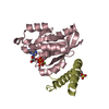
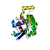
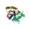
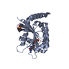
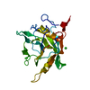
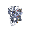
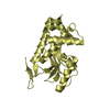
| Title | Crystal structure of a U4/U6 snRNP complex between human spliceosomal cyclophilin H and a U4/U6-60K peptide | ||||||
 Components Components |
| ||||||
 Keywords Keywords | ISOMERASE / cyclophilin / peptidyl-prolyl-cis/trans isomerase / spliceosome / snRNP / U4/U6-60K protein / WD protein | ||||||
| Function / homology |  Function and homology information Function and homology informationspliceosomal snRNP complex / U4/U6 snRNP / RNA splicing, via transesterification reactions / U2-type precatalytic spliceosome / U4 snRNA binding / cyclosporin A binding / U6 snRNA binding / ribonucleoprotein complex binding / Cajal body / RNA processing ...spliceosomal snRNP complex / U4/U6 snRNP / RNA splicing, via transesterification reactions / U2-type precatalytic spliceosome / U4 snRNA binding / cyclosporin A binding / U6 snRNA binding / ribonucleoprotein complex binding / Cajal body / RNA processing / positive regulation of viral genome replication / U4/U6 x U5 tri-snRNP complex / mRNA Splicing - Major Pathway / RNA splicing / peptidylprolyl isomerase / peptidyl-prolyl cis-trans isomerase activity / spliceosomal complex / mRNA splicing, via spliceosome / SARS-CoV-1 activates/modulates innate immune responses / protein folding / protein-containing complex assembly / nuclear speck / nucleoplasm / nucleus / cytoplasm / cytosol Similarity search - Function | ||||||
| Biological species |  Homo sapiens (human) Homo sapiens (human) | ||||||
| Method |  X-RAY DIFFRACTION / MOLECULAR REPLACEMENT / Resolution: 2 Å X-RAY DIFFRACTION / MOLECULAR REPLACEMENT / Resolution: 2 Å | ||||||
 Authors Authors | Reidt, U. / Wahl, M.C. / Horowitz, D.S. / Luehrmann, R. / Ficner, R. | ||||||
 Citation Citation | Journal: J.Mol.Biol. / Year: 2003 Title: Crystal structure of a complex between human spliceosomal cyclophilin H and a U4/U6 snRNP-60K peptide Authors: Reidt, U. / Wahl, M.C. / Fasshauer, D. / Horowitz, D.S. / Luehrmann, R. / Ficner, R. | ||||||
| History |
|
- Structure visualization
| Structure viewer | Molecule: MolmilJmol/JSmol |
|---|
- Downloads & links
-Download
| PDBx/mmCIF format | 1mzw.cif.gz | 57.6 KB | Display | PDBx/mmCIF format |
|---|---|---|---|---|
| PDB format | pdb1mzw.ent.gz | 41.3 KB | Display | PDB format |
| PDBx/mmJSON format | 1mzw.json.gz | Tree view | PDBx/mmJSON format | |
| Others |  Other downloads Other downloads |
-Validation report
| Arichive directory | https://data.pdbj.org/pub/pdb/validation_reports/mz/1mzwftp://data.pdbj.org/pub/pdb/validation_reports/mz/1mzw | HTTPS FTP |
|---|
-Related structure data
| Related structure data |  1qoiS S: Starting model for refinement |
|---|---|
| Similar structure data |


-Links
 PDBj
PDBj


- Assembly
| Deposited unit | 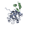
| ||||||||
|---|---|---|---|---|---|---|---|---|---|
| 1 |
| ||||||||
| Unit cell |
|
-Components
| #1: Protein | Mass: 19230.117 Da / Num. of mol.: 1 Source method: isolated from a genetically manipulated source Source: (gene. exp.) Homo sapiens (human) / Species (production host): Escherichia coli / Production host:  |
|---|---|
| #2: Protein/peptide | Mass: 3498.044 Da / Num. of mol.: 1 / Fragment: residues 107-137, internal domain / Source method: obtained synthetically / Details: peptide B106-B136 was chemically synthesized / References: UniProt: O43172 |
| #3: Water | ChemComp-HOH /  Mass: 18.015 Da / Num. of mol.: 260 / Source method: isolated from a natural source / Formula: H2O Mass: 18.015 Da / Num. of mol.: 260 / Source method: isolated from a natural source / Formula: H2O |
-Experimental details
-Experiment
| Experiment | Method: X-RAY DIFFRACTION / Number of used crystals: 1 |
|---|
- Sample preparation
| Crystal | Density Matthews: 2.56 Å3/Da / Density % sol: 51.88 % | ||||||||||||||||||||||||
|---|---|---|---|---|---|---|---|---|---|---|---|---|---|---|---|---|---|---|---|---|---|---|---|---|---|
| Crystal grow | Temperature: 294 K / Method: vapor diffusion, hanging drop / pH: 6.5 Details: PEG 8000, magnesium acetate, HEPES, pH 6.5, VAPOR DIFFUSION, HANGING DROP, temperature 294.0K | ||||||||||||||||||||||||
| Crystal grow | *PLUS Temperature: 21 ℃ / Method: vapor diffusion, hanging drop | ||||||||||||||||||||||||
| Components of the solutions | *PLUS
|
-Data collection
| Diffraction | Mean temperature: 100 K |
|---|---|
| Diffraction source | Source: ROTATING ANODE / Type: ENRAF-NONIUS FR591 / Wavelength: 1.5418 Å |
| Detector | Type: MARRESEARCH / Detector: IMAGE PLATE / Date: Nov 10, 2001 / Details: Osmic Mirrors |
| Radiation | Monochromator: Osmic Mirror / Protocol: SINGLE WAVELENGTH / Monochromatic (M) / Laue (L): M / Scattering type: x-ray |
| Radiation wavelength | Wavelength: 1.5418 Å / Relative weight: 1 |
| Reflection | Resolution: 2→100 Å / Num. obs: 55492 / % possible obs: 99.2 % / Observed criterion σ(F): 1 / Observed criterion σ(I): 1 / Rsym value: 0.063 |
| Reflection shell | Resolution: 2→2.1 Å / Rsym value: 0.236 / % possible all: 99 |
| Reflection | *PLUS Highest resolution: 2.1 Å / Num. obs: 13762 / Num. measured all: 55492 / Rmerge(I) obs: 0.063 |
| Reflection shell | *PLUS Highest resolution: 2.1 Å / Lowest resolution: 2.2 Å / % possible obs: 99 % / Rmerge(I) obs: 0.236 |
- Processing
| Software |
| |||||||||||||||||||||||||
|---|---|---|---|---|---|---|---|---|---|---|---|---|---|---|---|---|---|---|---|---|---|---|---|---|---|---|
| Refinement | Method to determine structure: MOLECULAR REPLACEMENT Starting model: PDB ENTRY 1QOI Resolution: 2→15 Å / Isotropic thermal model: ISOTROPIC / σ(F): 2 / σ(I): 2 / Stereochemistry target values: Engh & Huber
| |||||||||||||||||||||||||
| Refinement step | Cycle: LAST / Resolution: 2→15 Å
| |||||||||||||||||||||||||
| Refine LS restraints |
| |||||||||||||||||||||||||
| Refinement | *PLUS Highest resolution: 2.1 Å / % reflection Rfree: 10 % / Rfactor Rfree: 0.257 | |||||||||||||||||||||||||
| Solvent computation | *PLUS | |||||||||||||||||||||||||
| Displacement parameters | *PLUS | |||||||||||||||||||||||||
| LS refinement shell | *PLUS Rfactor Rfree: 0.275 / Rfactor Rwork: 0.212 |
