 Movie
Movie Controller

[English] 日本語
 Yorodumi
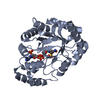
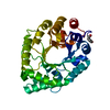
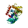
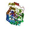
- PDB-1mzr: Structure of dkga from E.coli at 2.13 A resolution solved by mole... -
+ Open data

- Basic information
| Entry | Database: PDB / ID: 1mzr | ||||||
|---|---|---|---|---|---|---|---|
| Title | Structure of dkga from E.coli at 2.13 A resolution solved by molecular replacement | ||||||
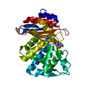
 Components Components | 2,5-diketo-D-gluconate reductase A | ||||||
 Keywords Keywords | OXIDOREDUCTASE / alpha/beta-barrel / aldo-ketoreductase / NADPH dependant / Bacterial targets at IGS-CNRS / France / BIGS / Structural Genomics | ||||||
| Function / homology |  Function and homology information Function and homology information2,5-didehydrogluconate reductase (2-dehydro-L-gulonate-forming) / 2,5-didehydrogluconate reductase activity / methylglyoxal catabolic process / alcohol dehydrogenase (NADP+) / methylglyoxal reductase (NADPH) (acetol producing) activity / alcohol dehydrogenase (NADP+) activity / Oxidoreductases; Acting on the CH-OH group of donors; With NAD+ or NADP+ as acceptor / aldose reductase (NADPH) activity / oxidoreductase activity, acting on the CH-OH group of donors, NAD or NADP as acceptor / cytosol Similarity search - Function | ||||||
| Biological species |  | ||||||
| Method |  X-RAY DIFFRACTION / SYNCHROTRON / MOLECULAR REPLACEMENT / Resolution: 2.13 Å X-RAY DIFFRACTION / SYNCHROTRON / MOLECULAR REPLACEMENT / Resolution: 2.13 Å | ||||||
 Authors Authors | Abergel, C. / Jeudy, S. / Monchois, V. / Claverie, J.M. / Bacterial targets at IGS-CNRS, France (BIGS) | ||||||
 Citation Citation | Journal: Proteins / Year: 2006 Title: Crystal structure of Escherichia coli DkgA, a broad-specificity aldo-keto reductase. Authors: Jeudy, S. / Monchois, V. / Maza, C. / Claverie, J.M. / Abergel, C. | ||||||
| History |
|
- Structure visualization
| Structure viewer | Molecule: MolmilJmol/JSmol |
|---|
- Downloads & links
-Download
| PDBx/mmCIF format | 1mzr.cif.gz | 134.8 KB | Display | PDBx/mmCIF format |
|---|---|---|---|---|
| PDB format | pdb1mzr.ent.gz | 104.3 KB | Display | PDB format |
| PDBx/mmJSON format | 1mzr.json.gz | Tree view | PDBx/mmJSON format | |
| Others |  Other downloads Other downloads |
-Validation report
| Arichive directory | https://data.pdbj.org/pub/pdb/validation_reports/mz/1mzrftp://data.pdbj.org/pub/pdb/validation_reports/mz/1mzr | HTTPS FTP |
|---|
-Related structure data
| Related structure data |  1a80S S: Starting model for refinement |
|---|---|
| Similar structure data | |
| Other databases |


-Links
 PDBj
PDBj
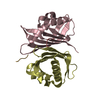
- Assembly
| Deposited unit | 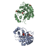
| ||||||||
|---|---|---|---|---|---|---|---|---|---|
| 1 | 
| ||||||||
| 2 | 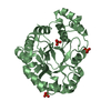
| ||||||||
| Unit cell |
| ||||||||
| Components on special symmetry positions |
|
-Components
| #1: Protein | Mass: 33686.398 Da / Num. of mol.: 2 Source method: isolated from a genetically manipulated source Source: (gene. exp.) References: UniProt: Q46857, Oxidoreductases; Acting on the CH-OH group of donors; With NAD+ or NADP+ as acceptor #2: Chemical | ChemComp-PO4 /   Mass: 94.971 Da / Num. of mol.: 5 / Source method: obtained synthetically / Formula: PO4 Mass: 94.971 Da / Num. of mol.: 5 / Source method: obtained synthetically / Formula: PO4#3: Chemical |   Mass: 92.094 Da / Num. of mol.: 2 / Source method: obtained synthetically / Formula: C3H8O3 Mass: 92.094 Da / Num. of mol.: 2 / Source method: obtained synthetically / Formula: C3H8O3#4: Water | ChemComp-HOH / |  Mass: 18.015 Da / Num. of mol.: 583 / Source method: isolated from a natural source / Formula: H2O Mass: 18.015 Da / Num. of mol.: 583 / Source method: isolated from a natural source / Formula: H2O |
|---|
-Experimental details
-Experiment
| Experiment | Method: X-RAY DIFFRACTION / Number of used crystals: 1 |
|---|
- Sample preparation
| Crystal | Density Matthews: 2.99 Å3/Da / Density % sol: 58.87 % | ||||||||||||||||||||||||||||||||||||||||||
|---|---|---|---|---|---|---|---|---|---|---|---|---|---|---|---|---|---|---|---|---|---|---|---|---|---|---|---|---|---|---|---|---|---|---|---|---|---|---|---|---|---|---|---|
| Crystal grow | Temperature: 293 K / Method: vapor diffusion, hanging drop / pH: 7 Details: potassium phosphate, sodium phosphate, hepes, glycerol, pH 7.0, VAPOR DIFFUSION, HANGING DROP, temperature 293K | ||||||||||||||||||||||||||||||||||||||||||
| Crystal grow | *PLUS pH: 8 / Method: vapor diffusion, sitting drop | ||||||||||||||||||||||||||||||||||||||||||
| Components of the solutions | *PLUS
|
-Data collection
| Diffraction | Mean temperature: 100 K |
|---|---|
| Diffraction source | Source: SYNCHROTRON / Site: ESRF  / Beamline: ID14-2 / Wavelength: 0.934 Å / Beamline: ID14-2 / Wavelength: 0.934 Å |
| Detector | Type: ADSC QUANTUM 4 / Detector: CCD / Date: Sep 15, 2001 |
| Radiation | Monochromator: monochromator 0.98 / Protocol: SINGLE WAVELENGTH / Monochromatic (M) / Laue (L): M / Scattering type: x-ray |
| Radiation wavelength | Wavelength: 0.934 Å / Relative weight: 1 |
| Reflection | Resolution: 2.13→37.1 Å / Num. all: 43958 / Num. obs: 43958 / % possible obs: 96.7 % / Observed criterion σ(F): 9 / Observed criterion σ(I): 3 / Redundancy: 4.9 % / Biso Wilson estimate: 26.751 Å2 / Rmerge(I) obs: 0.075 / Rsym value: 0.075 / Net I/σ(I): 8.6 |
| Reflection shell | Resolution: 2.13→2.2 Å / Redundancy: 4.8 % / Num. unique all: 4340 / Rsym value: 0.282 / % possible all: 98.3 |
| Reflection | *PLUS Highest resolution: 2.16 Å / Lowest resolution: 20 Å / Num. obs: 39423 |
| Reflection shell | *PLUS % possible obs: 96.7 % / Num. unique obs: 3932 / Rmerge(I) obs: 0.82 / Mean I/σ(I) obs: 2.6 |
- Processing
| Software |
| |||||||||||||||||||||||||
|---|---|---|---|---|---|---|---|---|---|---|---|---|---|---|---|---|---|---|---|---|---|---|---|---|---|---|
| Refinement | Method to determine structure: MOLECULAR REPLACEMENT Starting model: PDB entry 1A80 Resolution: 2.13→19.91 Å / Isotropic thermal model: RESTRAINED / Cross valid method: THROUGHOUT / σ(F): 0 / σ(I): 0 / Stereochemistry target values: Engh & Huber
| |||||||||||||||||||||||||
| Displacement parameters | Biso mean: 27 Å2
| |||||||||||||||||||||||||
| Refine analyze |
| |||||||||||||||||||||||||
| Refinement step | Cycle: LAST / Resolution: 2.13→19.91 Å
| |||||||||||||||||||||||||
| Refine LS restraints |
| |||||||||||||||||||||||||
| LS refinement shell | Resolution: 2.13→2.2 Å / Rfactor Rfree error: 0.01
| |||||||||||||||||||||||||
| Refinement | *PLUS Rfactor obs: 0.172 / Rfactor Rfree: 0.221 / Rfactor Rwork: 0.172 / Highest resolution: 2.16 Å / Lowest resolution: 20 Å | |||||||||||||||||||||||||
| Solvent computation | *PLUS | |||||||||||||||||||||||||
| Displacement parameters | *PLUS | |||||||||||||||||||||||||
| Refine LS restraints | *PLUS
|
