| 登録情報 | データベース: PDB / ID: 1mz9
|
|---|
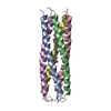
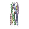
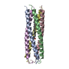
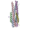
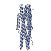
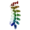
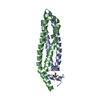
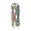
| タイトル | Storage function of COMP:the crystal structure of the coiled-coil domain in complex with vitamin D3 |
|---|
 要素 要素 | cartilage oligomeric matrix protein |
|---|
 キーワード キーワード | PROTEIN BINDING / pentameric coiled-coil domain |
|---|
| 機能・相同性 |  機能・相同性情報 機能・相同性情報
tendon development / negative regulation of hemostasis / cartilage homeostasis / ECM proteoglycans / vascular associated smooth muscle contraction / chondrocyte development / bone growth / vascular associated smooth muscle cell development / musculoskeletal movement / BMP binding ...tendon development / negative regulation of hemostasis / cartilage homeostasis / ECM proteoglycans / vascular associated smooth muscle contraction / chondrocyte development / bone growth / vascular associated smooth muscle cell development / musculoskeletal movement / BMP binding / chondrocyte proliferation / growth plate cartilage development / endochondral bone growth / Integrin cell surface interactions / positive regulation of chondrocyte proliferation / regulation of bone mineralization / muscle cell development / heparan sulfate proteoglycan binding / bone morphogenesis / cartilage development / collagen fibril organization / proteoglycan binding / limb development / artery morphogenesis / neuromuscular process / skin development / bone mineralization / protein secretion / response to unfolded protein / BMP signaling pathway / collagen binding / extracellular matrix / ossification / skeletal system development / protein homooligomerization / protein processing / platelet aggregation / multicellular organism growth / integrin binding / blood coagulation / cellular senescence / heparin binding / regulation of gene expression / protease binding / apoptotic process / calcium ion binding / negative regulation of apoptotic process / protein-containing complex / extracellular space / extracellular region類似検索 - 分子機能 Single alpha-helices involved in coiled-coils or other helix-helix interfaces - #10 / Thrombospondin/cartilage oligomeric matrix protein, coiled-coil domain / Thrombospondin-5, coiled coil region / : / Cartilage oligomeric matrix protein / Thrombospondin, C-terminal / Thrombospondin, type 3 repeat / Thrombospondin C-terminal region / Thrombospondin type-3 (TSP3) repeat profile. / Thrombospondin C-terminal domain profile. ...Single alpha-helices involved in coiled-coils or other helix-helix interfaces - #10 / Thrombospondin/cartilage oligomeric matrix protein, coiled-coil domain / Thrombospondin-5, coiled coil region / : / Cartilage oligomeric matrix protein / Thrombospondin, C-terminal / Thrombospondin, type 3 repeat / Thrombospondin C-terminal region / Thrombospondin type-3 (TSP3) repeat profile. / Thrombospondin C-terminal domain profile. / Thrombospondin, type 3-like repeat / Thrombospondin type 3 repeat / TSP type-3 repeat / : / Calcium-binding EGF domain / EGF-like calcium-binding, conserved site / Calcium-binding EGF-like domain signature. / EGF-like calcium-binding domain / Calcium-binding EGF-like domain / Epidermal growth factor-like domain. / EGF-like domain profile. / Growth factor receptor cysteine-rich domain superfamily / EGF-like domain signature 2. / Single alpha-helices involved in coiled-coils or other helix-helix interfaces / EGF-like domain / EF-hand calcium-binding domain. / Concanavalin A-like lectin/glucanase domain superfamily / Up-down Bundle / Mainly Alpha類似検索 - ドメイン・相同性 Chem-VDY / Cartilage oligomeric matrix protein類似検索 - 構成要素 |
|---|
| 生物種 |   Mus musculus (ハツカネズミ) Mus musculus (ハツカネズミ) |
|---|
| 手法 |  X線回折 / シンクロトロン / 分子置換 / 解像度: 1.7 Å X線回折 / シンクロトロン / 分子置換 / 解像度: 1.7 Å |
|---|
 データ登録者 データ登録者 | Stetefeld, J. |
|---|
 引用 引用 | ジャーナル: EMBO J. / 年: 2002
タイトル: Storage function of cartilage oligomeric matrix protein: the crystal structure of the coiled-coil domain in complex with vitamin D(3).
著者: Ozbek, S. / Engel, J. / Stetefeld, J. |
|---|
| 履歴 | | 登録 | 2002年10月7日 | 登録サイト: RCSB / 処理サイト: PDBJ |
|---|
| 改定 1.0 | 2003年1月28日 | Provider: repository / タイプ: Initial release |
|---|
| 改定 1.1 | 2008年4月28日 | Group: Version format compliance |
|---|
| 改定 1.2 | 2011年7月13日 | Group: Version format compliance |
|---|
| 改定 1.3 | 2022年12月21日 | Group: Data collection / Database references / Derived calculations
カテゴリ: database_2 / diffrn_source ...database_2 / diffrn_source / struct_ref_seq_dif / struct_site
Item: _database_2.pdbx_DOI / _database_2.pdbx_database_accession ..._database_2.pdbx_DOI / _database_2.pdbx_database_accession / _diffrn_source.pdbx_synchrotron_site / _struct_ref_seq_dif.details / _struct_site.pdbx_auth_asym_id / _struct_site.pdbx_auth_comp_id / _struct_site.pdbx_auth_seq_id |
|---|
| 改定 1.4 | 2024年11月20日 | Group: Data collection / Structure summary
カテゴリ: chem_comp_atom / chem_comp_bond ...chem_comp_atom / chem_comp_bond / pdbx_entry_details / pdbx_modification_feature |
|---|
|
|---|
 ムービー
ムービー コントローラー


 データを開く
 基本情報
基本情報 構造の表示
構造の表示 ダウンロードとリンク
 その他のダウンロード
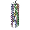
 PDBj
PDBj


 集合体
集合体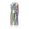

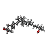
 分子量: 400.637 Da / 分子数: 2 / 由来タイプ: 合成 / 式: C27H44O2
 分子量: 18.015 Da / 分子数: 245 / 由来タイプ: 天然 / 式: H2O
 試料調製
試料調製 / ビームライン: BW7B
 解析
解析