 Movie
Movie Controller

[English] 日本語
 Yorodumi
- PDB-1mwu: Structure of methicillin acyl-Penicillin binding protein 2a from ... -
+ Open data

- Basic information
| Entry | Database: PDB / ID: 1mwu | ||||||
|---|---|---|---|---|---|---|---|
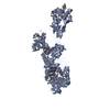
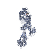
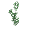
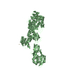
| Title | Structure of methicillin acyl-Penicillin binding protein 2a from methicillin resistant Staphylococcus aureus strain 27r at 2.60 A resolution. | ||||||
 Components Components | penicillin-binding protein 2a | ||||||
 Keywords Keywords | BIOSYNTHETIC PROTEIN / penicillin binding protein / beta-lactam / d / d-transpeptidase / d-carboxypeptidase / methicillin | ||||||
| Function / homology |  Function and homology information Function and homology informationpeptidoglycan glycosyltransferase / peptidoglycan L,D-transpeptidase activity / glycosyltransferase activity / penicillin binding / cell wall organization / response to antibiotic / plasma membrane Similarity search - Function | ||||||
| Biological species |   Staphylococcus aureus (bacteria) Staphylococcus aureus (bacteria) | ||||||
| Method |  X-RAY DIFFRACTION / Resolution: 2.6 Å X-RAY DIFFRACTION / Resolution: 2.6 Å | ||||||
 Authors Authors | Lim, D.C. / Strynadka, N.C.J. | ||||||
 Citation Citation | Journal: Nat.Struct.Biol. / Year: 2002 Title: Structural basis for the beta lactam resistance of PBP2a from methicillin-resistant Staphylococcus aureus. Authors: Lim, D. / Strynadka, N.C. | ||||||
| History |
| ||||||
| Remark 999 | SEQUENCE At the time of processing, the sequence for this strain was not present in a sequence ... SEQUENCE At the time of processing, the sequence for this strain was not present in a sequence database. However, the protein crystallized contains the engineered mutation Y23M and the first 22 residues were deleted. |
- Structure visualization
| Structure viewer | Molecule: MolmilJmol/JSmol |
|---|
- Downloads & links
-Download
| PDBx/mmCIF format | 1mwu.cif.gz | 259.5 KB | Display | PDBx/mmCIF format |
|---|---|---|---|---|
| PDB format | pdb1mwu.ent.gz | 208.4 KB | Display | PDB format |
| PDBx/mmJSON format | 1mwu.json.gz | Tree view | PDBx/mmJSON format | |
| Others |  Other downloads Other downloads |
-Validation report
| Arichive directory | https://data.pdbj.org/pub/pdb/validation_reports/mw/1mwuftp://data.pdbj.org/pub/pdb/validation_reports/mw/1mwu | HTTPS FTP |
|---|
-Related structure data


-Links
 PDBj
PDBj
- Assembly
| Deposited unit | 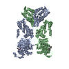
| ||||||||
|---|---|---|---|---|---|---|---|---|---|
| 1 | 
| ||||||||
| 2 | 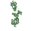
| ||||||||
| Unit cell |
|
-Components
| #1: Protein | Mass: 73777.250 Da / Num. of mol.: 2 / Fragment: residues 23-668 / Mutation: Y23M, delta 1-22 Source method: isolated from a genetically manipulated source Source: (gene. exp.) Staphylococcus aureus (bacteria) / Production host: #2: Chemical |   Mass: 382.431 Da / Num. of mol.: 2 / Source method: obtained synthetically / Formula: C17H22N2O6S / Comment: antibiotic*YM Mass: 382.431 Da / Num. of mol.: 2 / Source method: obtained synthetically / Formula: C17H22N2O6S / Comment: antibiotic*YM#3: Chemical | ChemComp-CD /   Mass: 112.411 Da / Num. of mol.: 5 / Source method: obtained synthetically / Formula: Cd Mass: 112.411 Da / Num. of mol.: 5 / Source method: obtained synthetically / Formula: Cd#4: Chemical | ChemComp-CL /   Mass: 35.453 Da / Num. of mol.: 4 / Source method: obtained synthetically / Formula: Cl Mass: 35.453 Da / Num. of mol.: 4 / Source method: obtained synthetically / Formula: Cl#5: Water | ChemComp-HOH / |  Mass: 18.015 Da / Num. of mol.: 67 / Source method: isolated from a natural source / Formula: H2O Mass: 18.015 Da / Num. of mol.: 67 / Source method: isolated from a natural source / Formula: H2OHas protein modification | Y | |
|---|
-Experimental details
-Experiment
| Experiment | Method: X-RAY DIFFRACTION / Number of used crystals: 1 |
|---|
- Sample preparation
| Crystal | Density Matthews: 2.62 Å3/Da / Density % sol: 53 % | ||||||||||||||||||||||||||||||||||||||||||
|---|---|---|---|---|---|---|---|---|---|---|---|---|---|---|---|---|---|---|---|---|---|---|---|---|---|---|---|---|---|---|---|---|---|---|---|---|---|---|---|---|---|---|---|
| Crystal grow | Temperature: 291 K / Method: vapor diffusion, hanging drop / pH: 7 Details: CdCl2, PEG550MME, HEPES, NaCl, pH 7, VAPOR DIFFUSION, HANGING DROP, temperature 291K | ||||||||||||||||||||||||||||||||||||||||||
| Crystal grow | *PLUS Temperature: 18 ℃ | ||||||||||||||||||||||||||||||||||||||||||
| Components of the solutions | *PLUS
|
-Data collection
| Diffraction | Mean temperature: 100 K |
|---|---|
| Diffraction source | Source: ROTATING ANODE / Type: RIGAKU RU200 / Wavelength: 1.54 Å |
| Detector | Type: MARRESEARCH / Detector: IMAGE PLATE / Date: Apr 13, 2002 / Details: Osmic |
| Radiation | Monochromator: Osmic / Protocol: SINGLE WAVELENGTH / Monochromatic (M) / Laue (L): M / Scattering type: x-ray |
| Radiation wavelength | Wavelength: 1.54 Å / Relative weight: 1 |
| Reflection | Resolution: 2.6→25 Å / Num. all: 47895 / Num. obs: 47895 / % possible obs: 98.1 % / Observed criterion σ(F): 0 / Observed criterion σ(I): 0 / Redundancy: 2.9 % / Biso Wilson estimate: 37.6 Å2 / Rsym value: 0.054 / Net I/σ(I): 12.7 |
| Reflection shell | Resolution: 2.6→2.7 Å / Mean I/σ(I) obs: 2 / Num. unique all: 4786 / Rsym value: 0.387 / % possible all: 99.7 |
| Reflection | *PLUS Lowest resolution: 25 Å / Rmerge(I) obs: 0.054 |
| Reflection shell | *PLUS % possible obs: 99.7 % / Rmerge(I) obs: 0.387 |
- Processing
| Software |
| ||||||||||||||||||||||||||||||||||||||||||||||||||||||||||||||||||||||||||||||||
|---|---|---|---|---|---|---|---|---|---|---|---|---|---|---|---|---|---|---|---|---|---|---|---|---|---|---|---|---|---|---|---|---|---|---|---|---|---|---|---|---|---|---|---|---|---|---|---|---|---|---|---|---|---|---|---|---|---|---|---|---|---|---|---|---|---|---|---|---|---|---|---|---|---|---|---|---|---|---|---|---|---|
| Refinement | Resolution: 2.6→24.68 Å / Rfactor Rfree error: 0.006 / Data cutoff high absF: 1943628.12 / Data cutoff low absF: 0 / Isotropic thermal model: RESTRAINED / Cross valid method: THROUGHOUT / σ(F): 0
| ||||||||||||||||||||||||||||||||||||||||||||||||||||||||||||||||||||||||||||||||
| Solvent computation | Solvent model: FLAT MODEL / Bsol: 30.3984 Å2 / ksol: 0.305723 e/Å3 | ||||||||||||||||||||||||||||||||||||||||||||||||||||||||||||||||||||||||||||||||
| Displacement parameters | Biso mean: 62.8 Å2
| ||||||||||||||||||||||||||||||||||||||||||||||||||||||||||||||||||||||||||||||||
| Refine analyze |
| ||||||||||||||||||||||||||||||||||||||||||||||||||||||||||||||||||||||||||||||||
| Refinement step | Cycle: LAST / Resolution: 2.6→24.68 Å
| ||||||||||||||||||||||||||||||||||||||||||||||||||||||||||||||||||||||||||||||||
| Refine LS restraints |
| ||||||||||||||||||||||||||||||||||||||||||||||||||||||||||||||||||||||||||||||||
| LS refinement shell | Resolution: 2.6→2.76 Å / Rfactor Rfree error: 0.023 / Total num. of bins used: 6
| ||||||||||||||||||||||||||||||||||||||||||||||||||||||||||||||||||||||||||||||||
| Xplor file |
| ||||||||||||||||||||||||||||||||||||||||||||||||||||||||||||||||||||||||||||||||
| Refinement | *PLUS Highest resolution: 2.6 Å / Lowest resolution: 25 Å / % reflection Rfree: 5 % / Rfactor obs: 0.242 / Rfactor Rfree: 0.302 / Rfactor Rwork: 0.242 | ||||||||||||||||||||||||||||||||||||||||||||||||||||||||||||||||||||||||||||||||
| Solvent computation | *PLUS | ||||||||||||||||||||||||||||||||||||||||||||||||||||||||||||||||||||||||||||||||
| Displacement parameters | *PLUS | ||||||||||||||||||||||||||||||||||||||||||||||||||||||||||||||||||||||||||||||||
| Refine LS restraints | *PLUS
| ||||||||||||||||||||||||||||||||||||||||||||||||||||||||||||||||||||||||||||||||
| LS refinement shell | *PLUS Rfactor Rfree: 0.441 / Rfactor Rwork: 0.374 |
