 Movie
Movie Controller

[English] 日本語
 Yorodumi
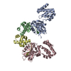
- PDB-1mw9: Crystal Structure of H365R mutant of 67 kDA N-terminal fragment o... -
+ Open data

- Basic information
| Entry | Database: PDB / ID: 1mw9 | ||||||
|---|---|---|---|---|---|---|---|
| Title | Crystal Structure of H365R mutant of 67 kDA N-terminal fragment of E. coli DNA Topoisomerase I | ||||||
 Components Components | DNA Topoisomerase I | ||||||
 Keywords Keywords | ISOMERASE / DNA TOPOISOMERASE / DECATENASE ENZYME / TOPRIM DOMAIN | ||||||
| Function / homology |  Function and homology information Function and homology informationRNA topoisomerase activity / DNA topoisomerase activity / DNA topoisomerase / DNA topoisomerase type I (single strand cut, ATP-independent) activity / DNA topological change / chromosome segregation / chromosome / DNA binding / zinc ion binding / cytosol Similarity search - Function | ||||||
| Biological species |  | ||||||
| Method |  X-RAY DIFFRACTION / SYNCHROTRON / MOLECULAR REPLACEMENT / Resolution: 1.67 Å X-RAY DIFFRACTION / SYNCHROTRON / MOLECULAR REPLACEMENT / Resolution: 1.67 Å | ||||||
 Authors Authors | Perry, K. / Mondragon, A. | ||||||
 Citation Citation | Journal: Structure / Year: 2003 Title: Structure of a Complex between E. coli DNA Topoisomerase I and Single-Stranded DNA. Authors: Perry, K. / Mondragon, A. #1: Journal: Nature / Year: 1994Title: Three-dimensional structure of the 67K N-terminal fragment of E. coli DNA topoisomerase I Authors: Lima, C.D. / Wang, J.C. / Mondragon, A. | ||||||
| History |
| ||||||
| Remark 999 | SEQUENCE THE AUTHOR'S SEQUENCE HAS ASN AT POSITION 549, WHICH HAS BEEN CONFIRMED BY SEQUENCING THE GENE. |
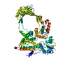
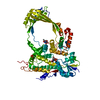
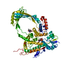
- Structure visualization
| Structure viewer | Molecule: MolmilJmol/JSmol |
|---|
- Downloads & links
-Download
| PDBx/mmCIF format | 1mw9.cif.gz | 138.8 KB | Display | PDBx/mmCIF format |
|---|---|---|---|---|
| PDB format | pdb1mw9.ent.gz | 108.8 KB | Display | PDB format |
| PDBx/mmJSON format | 1mw9.json.gz | Tree view | PDBx/mmJSON format | |
| Others |  Other downloads Other downloads |
-Validation report
| Arichive directory | https://data.pdbj.org/pub/pdb/validation_reports/mw/1mw9ftp://data.pdbj.org/pub/pdb/validation_reports/mw/1mw9 | HTTPS FTP |
|---|
-Related structure data
| Related structure data |  1mw8C  1eclS S: Starting model for refinement C: citing same article ( |
|---|---|
| Similar structure data |


-Links
 PDBj
PDBj


- Assembly
| Deposited unit | 
| ||||||||
|---|---|---|---|---|---|---|---|---|---|
| 1 |
| ||||||||
| Unit cell |
|
-Components
| #1: Protein | Mass: 66930.812 Da / Num. of mol.: 1 / Fragment: 67 kDa N-terminal fragment / Mutation: H365R Source method: isolated from a genetically manipulated source Source: (gene. exp.) | ||
|---|---|---|---|
| #2: Chemical | ChemComp-SO4 /   Mass: 96.063 Da / Num. of mol.: 9 / Source method: obtained synthetically / Formula: SO4 Mass: 96.063 Da / Num. of mol.: 9 / Source method: obtained synthetically / Formula: SO4#3: Water | ChemComp-HOH / |  Mass: 18.015 Da / Num. of mol.: 577 / Source method: isolated from a natural source / Formula: H2O Mass: 18.015 Da / Num. of mol.: 577 / Source method: isolated from a natural source / Formula: H2O |
-Experimental details
-Experiment
| Experiment | Method: X-RAY DIFFRACTION |
|---|
- Sample preparation
| Crystal | Density Matthews: 2.58 Å3/Da / Density % sol: 52.31 % |
|---|---|
| Crystal grow | Temperature: 287 K / Method: dialysis / pH: 8 / Details: ammonium sulfate, pH 8, DIALYSIS, temperature 287K |
| Crystal grow | *PLUS Method: vapor diffusion, hanging drop |
| Components of the solutions | *PLUS Conc.: 2.1 M / Common name: ammonium sulfate |
-Data collection
| Diffraction | Mean temperature: 103 K |
|---|---|
| Diffraction source | Source: SYNCHROTRON / Site: SSRL  / Beamline: BL9-1 / Wavelength: 0.98 Å / Beamline: BL9-1 / Wavelength: 0.98 Å |
| Detector | Type: MARRESEARCH / Detector: IMAGE PLATE / Date: Aug 15, 1999 |
| Radiation | Protocol: SINGLE WAVELENGTH / Monochromatic (M) / Laue (L): M / Scattering type: x-ray |
| Radiation wavelength | Wavelength: 0.98 Å / Relative weight: 1 |
| Reflection | Resolution: 1.67→50 Å / Num. all: 80202 / Num. obs: 79717 / % possible obs: 98.4 % / Observed criterion σ(I): 10737.6 / Biso Wilson estimate: 26 Å2 / Rmerge(I) obs: 0.066 / Rsym value: 0.055 / Net I/σ(I): 8.2 |
| Reflection | *PLUS Num. measured all: 398050 / Rmerge(I) obs: 0.057 |
| Reflection shell | *PLUS % possible obs: 98.4 % / Rmerge(I) obs: 0.407 |
- Processing
| Software |
| ||||||||||||||||||||||||||||||||||||||||||||||||||||||||||||||||||||||||||||||||||||||||||||||||||||||||||||||||||||||||||||||||||
|---|---|---|---|---|---|---|---|---|---|---|---|---|---|---|---|---|---|---|---|---|---|---|---|---|---|---|---|---|---|---|---|---|---|---|---|---|---|---|---|---|---|---|---|---|---|---|---|---|---|---|---|---|---|---|---|---|---|---|---|---|---|---|---|---|---|---|---|---|---|---|---|---|---|---|---|---|---|---|---|---|---|---|---|---|---|---|---|---|---|---|---|---|---|---|---|---|---|---|---|---|---|---|---|---|---|---|---|---|---|---|---|---|---|---|---|---|---|---|---|---|---|---|---|---|---|---|---|---|---|---|---|
| Refinement | Method to determine structure: MOLECULAR REPLACEMENT Starting model: PDB ENTRY 1ECL Resolution: 1.67→49.39 Å / Cor.coef. Fo:Fc: 0.954 / Cor.coef. Fo:Fc free: 0.952 / SU B: 2.785 / SU ML: 0.094 / Cross valid method: THROUGHOUT / ESU R: 0.11 / ESU R Free: 0.097 / Stereochemistry target values: MAXIMUM LIKELIHOOD / Details: HYDROGENS HAVE BEEN ADDED IN THE RIDING POSITIONS
| ||||||||||||||||||||||||||||||||||||||||||||||||||||||||||||||||||||||||||||||||||||||||||||||||||||||||||||||||||||||||||||||||||
| Solvent computation | Ion probe radii: 0.8 Å / Shrinkage radii: 0.8 Å / VDW probe radii: 1.4 Å / Solvent model: BABINET MODEL WITH MASK | ||||||||||||||||||||||||||||||||||||||||||||||||||||||||||||||||||||||||||||||||||||||||||||||||||||||||||||||||||||||||||||||||||
| Displacement parameters | Biso mean: 23.686 Å2
| ||||||||||||||||||||||||||||||||||||||||||||||||||||||||||||||||||||||||||||||||||||||||||||||||||||||||||||||||||||||||||||||||||
| Refinement step | Cycle: LAST / Resolution: 1.67→49.39 Å
| ||||||||||||||||||||||||||||||||||||||||||||||||||||||||||||||||||||||||||||||||||||||||||||||||||||||||||||||||||||||||||||||||||
| Refine LS restraints |
| ||||||||||||||||||||||||||||||||||||||||||||||||||||||||||||||||||||||||||||||||||||||||||||||||||||||||||||||||||||||||||||||||||
| LS refinement shell | Resolution: 1.67→1.713 Å / Rfactor Rfree error: 0.11 / Total num. of bins used: 20
| ||||||||||||||||||||||||||||||||||||||||||||||||||||||||||||||||||||||||||||||||||||||||||||||||||||||||||||||||||||||||||||||||||
| Software | *PLUS Version: 5 / Classification: refinement | ||||||||||||||||||||||||||||||||||||||||||||||||||||||||||||||||||||||||||||||||||||||||||||||||||||||||||||||||||||||||||||||||||
| Refinement | *PLUS Lowest resolution: 50 Å / Num. reflection obs: 75619 / Num. reflection Rfree: 4024 / Rfactor Rfree: 0.227 / Rfactor Rwork: 0.205 | ||||||||||||||||||||||||||||||||||||||||||||||||||||||||||||||||||||||||||||||||||||||||||||||||||||||||||||||||||||||||||||||||||
| Solvent computation | *PLUS | ||||||||||||||||||||||||||||||||||||||||||||||||||||||||||||||||||||||||||||||||||||||||||||||||||||||||||||||||||||||||||||||||||
| Displacement parameters | *PLUS | ||||||||||||||||||||||||||||||||||||||||||||||||||||||||||||||||||||||||||||||||||||||||||||||||||||||||||||||||||||||||||||||||||
| Refine LS restraints | *PLUS
|
