 Movie
Movie Controller

[English] 日本語
 Yorodumi
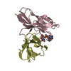
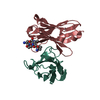
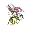
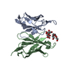
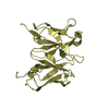
- PDB-1mvu: SINGLE CHAIN FV OF C219 HEAVY CHAIN V101L MUTANT IN COMPLEX WITH ... -
+ Open data

- Basic information
| Entry | Database: PDB / ID: 1mvu | ||||||
|---|---|---|---|---|---|---|---|
| Title | SINGLE CHAIN FV OF C219 HEAVY CHAIN V101L MUTANT IN COMPLEX WITH SYNTHETIC EPITOPE PEPTIDE | ||||||
 Components Components |
| ||||||
 Keywords Keywords | IMMUNE SYSTEM / SINGLE CHAIN FV / MONOCLONAL ANTIBODY / C219 / P-GLYCOPROTEIN / IMMUNOGLOBULIN / SITE-DIRECTED MUTAGENESIS | ||||||
| Function / homology |  Function and homology information Function and homology informationoligopeptide export from mitochondrion / terpenoid transport / ceramide floppase activity / floppase activity / phosphatidylethanolamine flippase activity / phosphatidylcholine floppase activity / ABC-type oligopeptide transporter activity / xenobiotic transport across blood-brain barrier / intercellular canaliculus / export across plasma membrane ...oligopeptide export from mitochondrion / terpenoid transport / ceramide floppase activity / floppase activity / phosphatidylethanolamine flippase activity / phosphatidylcholine floppase activity / ABC-type oligopeptide transporter activity / xenobiotic transport across blood-brain barrier / intercellular canaliculus / export across plasma membrane / P-type phospholipid transporter / ABC-type xenobiotic transporter / phospholipid translocation / ABC-type xenobiotic transporter activity / efflux transmembrane transporter activity / immunoglobulin complex / adaptive immune response / apical plasma membrane / mitochondrial inner membrane / ATP hydrolysis activity / extracellular region / ATP binding / cytoplasm Similarity search - Function | ||||||
| Biological species |  | ||||||
| Method |  X-RAY DIFFRACTION / MOLECULAR REPLACEMENT / Resolution: 1.78 Å X-RAY DIFFRACTION / MOLECULAR REPLACEMENT / Resolution: 1.78 Å | ||||||
 Authors Authors | Chan, D.C.M. / Kuntz, D.A. / Rose, D.R. | ||||||
 Citation Citation | Journal: To be Published Title: Single Chain C219 V(101H)L Mutant Antibody Complexed with a Helical Peptide Authors: Chan, D.C.M. / Hirama, T. / Mackenzie, C.R. / Kuntz, D.A. / Rose, D.R. #1: Journal: Proc.Natl.Acad.Sci.USA / Year: 1999Title: Antibody C19 Recognizes an Alpha-Helical Epitope on P-Glycoprotein Authors: Van den Elsen, J.M. / Kuntz, D.A. / Hoedemaeker, F.J. / Rose, D.R. #2: Journal: J.Biol.Chem. / Year: 1997Title: A Single Chain Fv Fragment of P-Glycoprotein-Specific Monoclonal Antibody C219. Design, Expression, and Crystal Structure at 2.4 A Resolution Authors: Hoedemaeker, F.J. / Signorelli, T. / Johns, K. / Kuntz, D.A. / Rose, D.R. | ||||||
| History |
| ||||||
| Remark 999 | SEQUENCE AT THE TIME OF PROCESSING, NO DATABASE REFERENCE SEQUENCE WAS AVAILABLE FOR C219 ANTIBODY ...SEQUENCE AT THE TIME OF PROCESSING, NO DATABASE REFERENCE SEQUENCE WAS AVAILABLE FOR C219 ANTIBODY HEAVY CHAIN. AUTHORS INFORMED THAT RESIDUE 101 OF THIS CHAIN WAS MUTATED FROM VAL TO LEU. |
- Structure visualization
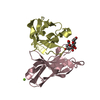
| Structure viewer | Molecule: MolmilJmol/JSmol |
|---|
- Downloads & links
-Download
| PDBx/mmCIF format | 1mvu.cif.gz | 65.8 KB | Display | PDBx/mmCIF format |
|---|---|---|---|---|
| PDB format | pdb1mvu.ent.gz | 47.7 KB | Display | PDB format |
| PDBx/mmJSON format | 1mvu.json.gz | Tree view | PDBx/mmJSON format | |
| Others |  Other downloads Other downloads |
-Validation report
| Arichive directory | https://data.pdbj.org/pub/pdb/validation_reports/mv/1mvuftp://data.pdbj.org/pub/pdb/validation_reports/mv/1mvu | HTTPS FTP |
|---|
-Related structure data
| Related structure data | |
|---|---|
| Similar structure data |


-Links
 PDBj
PDBj


- Assembly
| Deposited unit | 
| ||||||||
|---|---|---|---|---|---|---|---|---|---|
| 1 |
| ||||||||
| Unit cell |
|
-Components
| #1: Antibody | Mass: 12525.934 Da / Num. of mol.: 1 Source method: isolated from a genetically manipulated source Details: LIGHT AND HEAVY CHAINS LINKED WITH A SYNTHETIC (GGGGS)3 LINKER Source: (gene. exp.)  |
|---|---|
| #2: Antibody | Mass: 13442.889 Da / Num. of mol.: 1 Source method: isolated from a genetically manipulated source Details: LIGHT AND HEAVY CHAINS LINKED WITH A SYNTHETIC (GGGGS)3 LINKER Source: (gene. exp.) |
| #3: Protein/peptide | Mass: 1473.654 Da / Num. of mol.: 1 / Fragment: ATP-binding domain (Residues 1-13) / Source method: obtained synthetically Details: THE PEPTIDE WAS CHEMICALLY SYNTHESIZED. THE SEQUENCE OF THE PEPTIDE IS NATURALLY FOUND IN CRICETULUS GRISEUS (CHINESE HAMSTER). References: GenBank: 191155, UniProt: P21448*PLUS |
| #4: Chemical | ChemComp-SO4 /   Mass: 96.063 Da / Num. of mol.: 1 / Source method: obtained synthetically / Formula: SO4 Mass: 96.063 Da / Num. of mol.: 1 / Source method: obtained synthetically / Formula: SO4 |
| #5: Water | ChemComp-HOH /  Mass: 18.015 Da / Num. of mol.: 190 / Source method: isolated from a natural source / Formula: H2O Mass: 18.015 Da / Num. of mol.: 190 / Source method: isolated from a natural source / Formula: H2O |
| Has protein modification | Y |
-Experimental details
-Experiment
| Experiment | Method: X-RAY DIFFRACTION / Number of used crystals: 1 |
|---|
- Sample preparation
| Crystal | Density Matthews: 2.27 Å3/Da / Density % sol: 45.72 % |
|---|---|
| Crystal grow | Temperature: 298 K / Method: vapor diffusion, hanging drop / pH: 9 Details: PEG8000, lithium sulfate, pH 9, VAPOR DIFFUSION, HANGING DROP, temperature 298K |
-Data collection
| Diffraction | Mean temperature: 100 K |
|---|---|
| Diffraction source | Source: ROTATING ANODE / Type: RIGAKU / Wavelength: 1.5418 Å |
| Detector | Type: RIGAKU / Detector: IMAGE PLATE / Date: Jun 10, 2002 / Details: mirrors |
| Radiation | Monochromator: Quartz crystal / Protocol: SINGLE WAVELENGTH / Monochromatic (M) / Laue (L): M / Scattering type: x-ray |
| Radiation wavelength | Wavelength: 1.5418 Å / Relative weight: 1 |
| Reflection | Resolution: 1.78→20 Å / Num. obs: 23268 / % possible obs: 98.3 % / Observed criterion σ(F): 2 / Observed criterion σ(I): 3 / Biso Wilson estimate: 17 Å2 |
| Reflection shell | Resolution: 1.78→1.84 Å / % possible all: 96.9 |
- Processing
| Software |
| ||||||||||||||||||||||||||||||||||||||||||||||||||||||||||||
|---|---|---|---|---|---|---|---|---|---|---|---|---|---|---|---|---|---|---|---|---|---|---|---|---|---|---|---|---|---|---|---|---|---|---|---|---|---|---|---|---|---|---|---|---|---|---|---|---|---|---|---|---|---|---|---|---|---|---|---|---|---|
| Refinement | Method to determine structure: MOLECULAR REPLACEMENT / Resolution: 1.78→19.27 Å / Rfactor Rfree error: 0.007 / Data cutoff high absF: 385673.56 / Data cutoff high rms absF: 385673.56 / Data cutoff low absF: 0 / Isotropic thermal model: RESTRAINED / Cross valid method: THROUGHOUT / σ(F): 0 / Stereochemistry target values: Engh & Huber
| ||||||||||||||||||||||||||||||||||||||||||||||||||||||||||||
| Solvent computation | Solvent model: FLAT MODEL / Bsol: 47.8686 Å2 / ksol: 0.402656 e/Å3 | ||||||||||||||||||||||||||||||||||||||||||||||||||||||||||||
| Displacement parameters | Biso mean: 23.4 Å2
| ||||||||||||||||||||||||||||||||||||||||||||||||||||||||||||
| Refine analyze |
| ||||||||||||||||||||||||||||||||||||||||||||||||||||||||||||
| Refinement step | Cycle: LAST / Resolution: 1.78→19.27 Å
| ||||||||||||||||||||||||||||||||||||||||||||||||||||||||||||
| Refine LS restraints |
| ||||||||||||||||||||||||||||||||||||||||||||||||||||||||||||
| LS refinement shell | Resolution: 1.78→1.89 Å / Rfactor Rfree error: 0.023 / Total num. of bins used: 6
| ||||||||||||||||||||||||||||||||||||||||||||||||||||||||||||
| Xplor file |
|
