 Movie
Movie Controller

[English] 日本語
 Yorodumi
- PDB-1muq: X-ray Crystal Structure of Rattlesnake Venom Complexed With Thiod... -
+ Open data

- Basic information
| Entry | Database: PDB / ID: 1muq | ||||||||||||
|---|---|---|---|---|---|---|---|---|---|---|---|---|---|
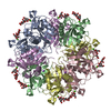
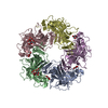
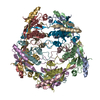
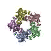
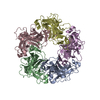
| Title | X-ray Crystal Structure of Rattlesnake Venom Complexed With Thiodigalactoside | ||||||||||||
 Components Components | Galactose-specific lectin | ||||||||||||
 Keywords Keywords | SUGAR BINDING PROTEIN / C-type lectin / protein-carbohydrate complex / decamer / calcium binding | ||||||||||||
| Function / homology |  Function and homology information Function and homology information | ||||||||||||
| Biological species |  Crotalus atrox (western diamondback rattlesnake) Crotalus atrox (western diamondback rattlesnake) | ||||||||||||
| Method |  X-RAY DIFFRACTION / SYNCHROTRON / MIR / Resolution: 2.3 Å X-RAY DIFFRACTION / SYNCHROTRON / MIR / Resolution: 2.3 Å | ||||||||||||
 Authors Authors | Walker, J.R. / Nagar, B. / Young, N.M. / Hirama, T. / Rini, J.M. | ||||||||||||
 Citation Citation | Journal: Biochemistry / Year: 2004 Title: X-ray Crystal Structure of a Galactose-Specific C-Type Lectin Possessing a Novel Decameric Quaternary Structure. Authors: Walker, J.R. / Nagar, B. / Young, N.M. / Hirama, T. / Rini, J.M. #1: Journal: Biochem.J. / Year: 1984Title: Isolation and characterization of three Ca2+-dependent beta-galactoside-specific lectins from snake venoms Authors: Gartner, T.K. / Ogilvie, M.L. #2: Journal: J.Biol.Chem. / Year: 1991Title: Complete primary structure of a galactose-specific lectin from the venom of the rattlesnake Crotalus atrox. Homologies with Ca2(+)-dependent-type lectins Authors: Hirabayashi, J.T. / Kusunoki, T. / Kasai, K. | ||||||||||||
| History |
|
- Structure visualization
| Structure viewer | Molecule: MolmilJmol/JSmol |
|---|
- Downloads & links
-Download
| PDBx/mmCIF format | 1muq.cif.gz | 159.7 KB | Display | PDBx/mmCIF format |
|---|---|---|---|---|
| PDB format | pdb1muq.ent.gz | 126.5 KB | Display | PDB format |
| PDBx/mmJSON format | 1muq.json.gz | Tree view | PDBx/mmJSON format | |
| Others |  Other downloads Other downloads |
-Validation report
| Arichive directory | https://data.pdbj.org/pub/pdb/validation_reports/mu/1muqftp://data.pdbj.org/pub/pdb/validation_reports/mu/1muq | HTTPS FTP |
|---|
-Related structure data


-Links
 PDBj
PDBj


- Assembly
| Deposited unit | 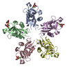
| ||||||||
|---|---|---|---|---|---|---|---|---|---|
| 1 | 
| ||||||||
| 2 |
| ||||||||
| Unit cell |
| ||||||||
| Details | The biological assembly is a decamer generated from the pentamer in the asymetric unit by the two fold axis -x, -y, z+1/2 |
-Components
-Protein , 1 types, 5 molecules ABCDE
| #1: Protein | Mass: 16312.377 Da / Num. of mol.: 5 / Source method: isolated from a natural source / Details: Isolated from venom Source: (natural) Crotalus atrox (western diamondback rattlesnake)References: UniProt: P21963 |
|---|
-Sugars , 2 types, 4 molecules 
| #2: Polysaccharide | 1-thio-beta-D-galactopyranose-(1-1)-beta-D-galactopyranose  Type: oligosaccharide, Oligosaccharide / Class: Substrate analog / Mass: 358.362 Da / Num. of mol.: 1 Type: oligosaccharide, Oligosaccharide / Class: Substrate analog / Mass: 358.362 Da / Num. of mol.: 1Source method: isolated from a genetically manipulated source Details: oligosaccharide with S-glycosidic bond between monosaccharides, and with reducing-end-to-reducing-end glycosidic bond References: thiodigalactoside |
|---|---|
| #5: Sugar |  Type: D-saccharide, beta linking / Mass: 180.156 Da / Num. of mol.: 3 Type: D-saccharide, beta linking / Mass: 180.156 Da / Num. of mol.: 3Source method: isolated from a genetically manipulated source Formula: C6H12O6 |
-Non-polymers , 3 types, 161 molecules 


| #3: Chemical | ChemComp-CA /  Mass: 40.078 Da / Num. of mol.: 5 / Source method: obtained synthetically / Formula: Ca Mass: 40.078 Da / Num. of mol.: 5 / Source method: obtained synthetically / Formula: Ca#4: Chemical | ChemComp-NA /  Mass: 22.990 Da / Num. of mol.: 5 / Source method: obtained synthetically / Formula: Na Mass: 22.990 Da / Num. of mol.: 5 / Source method: obtained synthetically / Formula: Na#6: Water | ChemComp-HOH / | Mass: 18.015 Da / Num. of mol.: 151 / Source method: isolated from a natural source / Formula: H2O |
|---|
-Details
| Has protein modification | Y |
|---|
-Experimental details
-Experiment
| Experiment | Method: X-RAY DIFFRACTION / Number of used crystals: 1 |
|---|
- Sample preparation
| Crystal | Density Matthews: 2.43 Å3/Da / Density % sol: 49.42 % | ||||||||||||||||||||||||||||||||||||||||||||||||||||||||
|---|---|---|---|---|---|---|---|---|---|---|---|---|---|---|---|---|---|---|---|---|---|---|---|---|---|---|---|---|---|---|---|---|---|---|---|---|---|---|---|---|---|---|---|---|---|---|---|---|---|---|---|---|---|---|---|---|---|
| Crystal grow | Temperature: 278 K / Method: vapor diffusion, hanging drop / pH: 7 Details: sodium formate, glycerol, hepes, pH 7.0, VAPOR DIFFUSION, HANGING DROP, temperature 278K | ||||||||||||||||||||||||||||||||||||||||||||||||||||||||
| Crystal grow | *PLUS Temperature: 4 ℃ / pH: 8 / Method: vapor diffusion, hanging drop | ||||||||||||||||||||||||||||||||||||||||||||||||||||||||
| Components of the solutions | *PLUS
|
-Data collection
| Diffraction | Mean temperature: 100 K |
|---|---|
| Diffraction source | Source: SYNCHROTRON / Site: CHESS  / Beamline: A1 / Wavelength: 0.909 Å / Beamline: A1 / Wavelength: 0.909 Å |
| Detector | Type: ADSC QUANTUM 4 / Detector: CCD / Date: Jun 10, 1998 |
| Radiation | Monochromator: horizontally focussing, 5.05 degrees asymmetric cut Si(111) Protocol: SINGLE WAVELENGTH / Monochromatic (M) / Laue (L): M / Scattering type: x-ray |
| Radiation wavelength | Wavelength: 0.909 Å / Relative weight: 1 |
| Reflection | Resolution: 2.2→35.13 Å / Num. all: 40555 / Num. obs: 40555 / % possible obs: 94.8 % / Observed criterion σ(F): 0 / Observed criterion σ(I): -3 / Redundancy: 14.6 % / Biso Wilson estimate: 29.9 Å2 / Rmerge(I) obs: 0.109 / Net I/σ(I): 12.37 |
| Reflection shell | Resolution: 2.2→2.24 Å / Redundancy: 2.3 % / Rmerge(I) obs: 0.4 / Mean I/σ(I) obs: 2.42 / Num. unique all: 1653 / % possible all: 82.9 |
| Reflection | *PLUS Highest resolution: 2.2 Å / Lowest resolution: 40 Å / Num. obs: 33477 |
| Reflection shell | *PLUS % possible obs: 82.9 % / Rmerge(I) obs: 0.402 |
- Processing
| Software |
| ||||||||||||||||||||||||||||||||||||
|---|---|---|---|---|---|---|---|---|---|---|---|---|---|---|---|---|---|---|---|---|---|---|---|---|---|---|---|---|---|---|---|---|---|---|---|---|---|
| Refinement | Method to determine structure: MIR / Resolution: 2.3→35.13 Å / Rfactor Rfree error: 0.004 / Data cutoff high absF: 3277247.32 / Data cutoff high rms absF: 3277247.32 / Data cutoff low absF: 0 / Isotropic thermal model: RESTRAINED / Cross valid method: THROUGHOUT / σ(F): 0 / Stereochemistry target values: Engh & Huber Details: Positions of sulfer atoms contributing to the disulfide bond between pentamers were fixed during refinement.
| ||||||||||||||||||||||||||||||||||||
| Solvent computation | Solvent model: FLAT MODEL / Bsol: 48.2996 Å2 / ksol: 0.439546 e/Å3 | ||||||||||||||||||||||||||||||||||||
| Displacement parameters | Biso mean: 48.6 Å2
| ||||||||||||||||||||||||||||||||||||
| Refine analyze |
| ||||||||||||||||||||||||||||||||||||
| Refinement step | Cycle: LAST / Resolution: 2.3→35.13 Å
| ||||||||||||||||||||||||||||||||||||
| Refine LS restraints |
| ||||||||||||||||||||||||||||||||||||
| LS refinement shell | Resolution: 2.3→2.44 Å / Rfactor Rfree error: 0.014 / Total num. of bins used: 6
| ||||||||||||||||||||||||||||||||||||
| Xplor file |
| ||||||||||||||||||||||||||||||||||||
| Refinement | *PLUS Highest resolution: 2.3 Å / Lowest resolution: 35 Å / Rfactor Rfree: 0.26 | ||||||||||||||||||||||||||||||||||||
| Solvent computation | *PLUS | ||||||||||||||||||||||||||||||||||||
| Displacement parameters | *PLUS | ||||||||||||||||||||||||||||||||||||
| Refine LS restraints | *PLUS
|
