 Movie
Movie Controller


+ Open data

- Basic information
| Entry | Database: PDB / ID: 3cae | ||||||
|---|---|---|---|---|---|---|---|
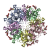
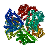
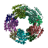
| Title | Structure of NNQQNY as an insert in T7 endonuclease I | ||||||
 Components Components | Endonuclease I | ||||||
 Keywords Keywords | HYDROLASE / T7 endonuclease I / amyloid / steric zipper | ||||||
| Function / homology |  Function and homology information Function and homology informationdegradation of host chromosome by virus / deoxyribonuclease IV / deoxyribonuclease IV (phage-T4-induced) activity / double-stranded DNA endonuclease activity / crossover junction DNA endonuclease activity / DNA integration / symbiont-mediated suppression of host gene expression / DNA binding Similarity search - Function | ||||||
| Biological species |   Enterobacteria phage T7 (virus) Enterobacteria phage T7 (virus) | ||||||
| Method |  X-RAY DIFFRACTION / SYNCHROTRON / MOLECULAR REPLACEMENT / Resolution: 3 Å X-RAY DIFFRACTION / SYNCHROTRON / MOLECULAR REPLACEMENT / Resolution: 3 Å | ||||||
 Authors Authors | Guo, Z. / Eisenberg, D. | ||||||
 Citation Citation | Journal: Protein Sci. / Year: 2008 Title: The structure of a fibril-forming sequence, NNQQNY, in the context of a globular fold. Authors: Guo, Z. / Eisenberg, D. | ||||||
| History |
|
- Structure visualization
| Structure viewer | Molecule: MolmilJmol/JSmol |
|---|
- Downloads & links
-Download
| PDBx/mmCIF format | 3cae.cif.gz | 277 KB | Display | PDBx/mmCIF format |
|---|---|---|---|---|
| PDB format | pdb3cae.ent.gz | 226.1 KB | Display | PDB format |
| PDBx/mmJSON format | 3cae.json.gz | Tree view | PDBx/mmJSON format | |
| Others |  Other downloads Other downloads |
-Validation report
| Arichive directory | https://data.pdbj.org/pub/pdb/validation_reports/ca/3caeftp://data.pdbj.org/pub/pdb/validation_reports/ca/3cae | HTTPS FTP |
|---|
-Related structure data
| Related structure data |  1fzrS S: Starting model for refinement |
|---|---|
| Similar structure data |


-Links
 PDBj
PDBj


- Assembly
| Deposited unit | 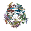
| ||||||||
|---|---|---|---|---|---|---|---|---|---|
| 1 |
| ||||||||
| Unit cell |
|
-Components
| #1: Protein | Mass: 15650.829 Da / Num. of mol.: 10 Source method: isolated from a genetically manipulated source Source: (gene. exp.) Enterobacteria phage T7 (virus) / Production host:  #2: Water | ChemComp-HOH / |  Mass: 18.015 Da / Num. of mol.: 15 / Source method: isolated from a natural source / Formula: H2O Mass: 18.015 Da / Num. of mol.: 15 / Source method: isolated from a natural source / Formula: H2O |
|---|
-Experimental details
-Experiment
| Experiment | Method: X-RAY DIFFRACTION / Number of used crystals: 1 |
|---|
- Sample preparation
| Crystal | Density Matthews: 3.5 Å3/Da / Density % sol: 64.88 % |
|---|---|
| Crystal grow | Temperature: 293 K / Method: vapor diffusion, hanging drop / pH: 5.5 Details: 2.6 M ammonium sulfate, 0.1 M sodium citrate, 2% dimethyl formamide, pH 5.5, VAPOR DIFFUSION, HANGING DROP, temperature 293K |
-Data collection
| Diffraction | Mean temperature: 100 K |
|---|---|
| Diffraction source | Source: SYNCHROTRON / Site: ALS  / Beamline: 8.2.2 / Wavelength: 1 Å / Beamline: 8.2.2 / Wavelength: 1 Å |
| Detector | Type: ADSC QUANTUM 315 / Detector: CCD / Date: Mar 1, 2007 |
| Radiation | Monochromator: Double Crystal Si(111) / Protocol: SINGLE WAVELENGTH / Monochromatic (M) / Laue (L): M / Scattering type: x-ray |
| Radiation wavelength | Wavelength: 1 Å / Relative weight: 1 |
| Reflection | Resolution: 3→90 Å / Num. all: 43123 / Num. obs: 43123 / % possible obs: 99.4 % / Observed criterion σ(F): 0 / Observed criterion σ(I): 0 / Redundancy: 3.2 % / Rsym value: 0.052 / Χ2: 1 / Net I/σ(I): 14.2 |
| Reflection shell | Resolution: 3→3.11 Å / Redundancy: 3 % / Mean I/σ(I) obs: 1.6 / Num. unique all: 4219 / Rsym value: 0.429 / Χ2: 1.006 / % possible all: 98.3 |
- Processing
| Software |
| ||||||||||||||||||||||||||||
|---|---|---|---|---|---|---|---|---|---|---|---|---|---|---|---|---|---|---|---|---|---|---|---|---|---|---|---|---|---|
| Refinement | Method to determine structure: MOLECULAR REPLACEMENT Starting model: PDB entry 1FZR, residues 18-43 of molecule A and residues 51-145 of molecule B Resolution: 3→90 Å / Cross valid method: FREE R / σ(F): 0 / Stereochemistry target values: Engh & Huber
| ||||||||||||||||||||||||||||
| Solvent computation | Bsol: 62.292 Å2 | ||||||||||||||||||||||||||||
| Displacement parameters | Biso mean: 86.205 Å2
| ||||||||||||||||||||||||||||
| Refinement step | Cycle: LAST / Resolution: 3→90 Å
| ||||||||||||||||||||||||||||
| Refine LS restraints |
| ||||||||||||||||||||||||||||
| Xplor file |
|
