 Movie
Movie Controller

[English] 日本語
 Yorodumi
- PDB-4a34: Crystal structure of the fucose mutarotase in complex with L-fuco... -
+ Open data

- Basic information
| Entry | Database: PDB / ID: 4a34 | ||||||
|---|---|---|---|---|---|---|---|
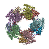
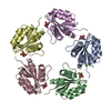
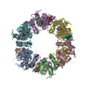
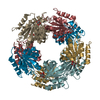
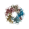
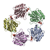
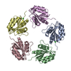
| Title | Crystal structure of the fucose mutarotase in complex with L-fucose from Streptococcus pneumoniae | ||||||
 Components Components | RBSD/FUCU TRANSPORT PROTEIN FAMILY PROTEIN | ||||||
 Keywords Keywords | ISOMERASE | ||||||
| Function / homology |  Function and homology information Function and homology informationL-fucose mutarotase activity / D-ribose pyranase / D-ribose pyranase activity / monosaccharide metabolic process / fucose binding / fucose metabolic process / monosaccharide binding Similarity search - Function | ||||||
| Biological species |   STREPTOCOCCUS PNEUMONIAE (bacteria) STREPTOCOCCUS PNEUMONIAE (bacteria) | ||||||
| Method |  X-RAY DIFFRACTION / OTHER / Resolution: 2.5 Å X-RAY DIFFRACTION / OTHER / Resolution: 2.5 Å | ||||||
 Authors Authors | Higgins, M.A. / Boraston, A.B. | ||||||
 Citation Citation | Journal: Acta Crystallogr.,Sect.F / Year: 2011 Title: Structure of the Fucose Mutarotase from Streptococcus Pneumoniae in Complex with L-Fucose Authors: Higgins, M.A. / Boraston, A.B. | ||||||
| History |
|
- Structure visualization
| Structure viewer | Molecule: MolmilJmol/JSmol |
|---|
- Downloads & links
-Download
| PDBx/mmCIF format | 4a34.cif.gz | 544.7 KB | Display | PDBx/mmCIF format |
|---|---|---|---|---|
| PDB format | pdb4a34.ent.gz | 454 KB | Display | PDB format |
| PDBx/mmJSON format | 4a34.json.gz | Tree view | PDBx/mmJSON format | |
| Others |  Other downloads Other downloads |
-Validation report
| Arichive directory | https://data.pdbj.org/pub/pdb/validation_reports/a3/4a34ftp://data.pdbj.org/pub/pdb/validation_reports/a3/4a34 | HTTPS FTP |
|---|
-Related structure data
| Similar structure data |
|---|

-Links
 PDBj
PDBj- Assembly
| Deposited unit | 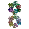
| ||||||||
|---|---|---|---|---|---|---|---|---|---|
| 1 | 
| ||||||||
| 2 | 
| ||||||||
| Unit cell |
|
-Components
| #1: Protein | Mass: 16574.094 Da / Num. of mol.: 20 Source method: isolated from a genetically manipulated source Source: (gene. exp.) STREPTOCOCCUS PNEUMONIAE (bacteria) / Strain: TIGR4 / Production host: #2: Sugar | ChemComp-FUL /   Type: L-saccharide, beta linking / Mass: 164.156 Da / Num. of mol.: 20 Type: L-saccharide, beta linking / Mass: 164.156 Da / Num. of mol.: 20Source method: isolated from a genetically manipulated source Formula: C6H12O5 #3: Chemical | ChemComp-K /   Mass: 39.098 Da / Num. of mol.: 10 / Source method: obtained synthetically / Formula: K Mass: 39.098 Da / Num. of mol.: 10 / Source method: obtained synthetically / Formula: K#4: Water | ChemComp-HOH / |  Mass: 18.015 Da / Num. of mol.: 850 / Source method: isolated from a natural source / Formula: H2O Mass: 18.015 Da / Num. of mol.: 850 / Source method: isolated from a natural source / Formula: H2O |
|---|
-Experimental details
-Experiment
| Experiment | Method: X-RAY DIFFRACTION |
|---|
- Sample preparation
| Crystal | Density Matthews: 2.29 Å3/Da / Density % sol: 46.39 % / Description: NONE |
|---|
-Data collection
| Diffraction | Mean temperature: 113 K |
|---|---|
| Diffraction source | Source: ROTATING ANODE / Wavelength: 1.5418 |
| Detector | Type: RIGAKU AREA DETECTOR / Details: OSMIC 'BLUE' |
| Radiation | Protocol: SINGLE WAVELENGTH / Monochromatic (M) / Laue (L): M / Scattering type: x-ray |
| Radiation wavelength | Wavelength: 1.5418 Å / Relative weight: 1 |
| Reflection | Resolution: 2.5→20 Å / Num. obs: 97486 / % possible obs: 99.7 % / Observed criterion σ(I): 2 / Redundancy: 4 % / Biso Wilson estimate: 38.9 Å2 / Rmerge(I) obs: 0.09 |
- Processing
| Software | Name: REFMAC / Version: 5.5.0109 / Classification: refinement | ||||||||||||||||||||||||||||||||||||||||||||||||||||||||||||||||||||||||||||||||||||||||||||||||||||||||||||||||||||||||||||||||||||||||||||||||||||||||||||||||||||||||||||||||||||||
|---|---|---|---|---|---|---|---|---|---|---|---|---|---|---|---|---|---|---|---|---|---|---|---|---|---|---|---|---|---|---|---|---|---|---|---|---|---|---|---|---|---|---|---|---|---|---|---|---|---|---|---|---|---|---|---|---|---|---|---|---|---|---|---|---|---|---|---|---|---|---|---|---|---|---|---|---|---|---|---|---|---|---|---|---|---|---|---|---|---|---|---|---|---|---|---|---|---|---|---|---|---|---|---|---|---|---|---|---|---|---|---|---|---|---|---|---|---|---|---|---|---|---|---|---|---|---|---|---|---|---|---|---|---|---|---|---|---|---|---|---|---|---|---|---|---|---|---|---|---|---|---|---|---|---|---|---|---|---|---|---|---|---|---|---|---|---|---|---|---|---|---|---|---|---|---|---|---|---|---|---|---|---|---|
| Refinement | Method to determine structure: OTHER Starting model: NONE Resolution: 2.5→19.97 Å / Cor.coef. Fo:Fc: 0.935 / Cor.coef. Fo:Fc free: 0.884 / SU B: 13.176 / SU ML: 0.286 / Cross valid method: THROUGHOUT / ESU R: 1.081 / ESU R Free: 0.359 / Stereochemistry target values: MAXIMUM LIKELIHOOD Details: HYDROGENS HAVE BEEN ADDED IN THE RIDING POSITIONS. U VALUES REFINED INDIVIDUALLY.
| ||||||||||||||||||||||||||||||||||||||||||||||||||||||||||||||||||||||||||||||||||||||||||||||||||||||||||||||||||||||||||||||||||||||||||||||||||||||||||||||||||||||||||||||||||||||
| Solvent computation | Ion probe radii: 0.8 Å / Shrinkage radii: 0.8 Å / VDW probe radii: 1.4 Å / Solvent model: MASK | ||||||||||||||||||||||||||||||||||||||||||||||||||||||||||||||||||||||||||||||||||||||||||||||||||||||||||||||||||||||||||||||||||||||||||||||||||||||||||||||||||||||||||||||||||||||
| Displacement parameters | Biso mean: 38.226 Å2
| ||||||||||||||||||||||||||||||||||||||||||||||||||||||||||||||||||||||||||||||||||||||||||||||||||||||||||||||||||||||||||||||||||||||||||||||||||||||||||||||||||||||||||||||||||||||
| Refinement step | Cycle: LAST / Resolution: 2.5→19.97 Å
| ||||||||||||||||||||||||||||||||||||||||||||||||||||||||||||||||||||||||||||||||||||||||||||||||||||||||||||||||||||||||||||||||||||||||||||||||||||||||||||||||||||||||||||||||||||||
| Refine LS restraints |
|