[English] 日本語
Yorodumi
- PDB-1mu5: Structure of topoisomerase subunit -

+
Open data


ID or keywords:

Loading...

-
Basic information

Entry
Database: PDB / ID: 1mu5
TitleStructure of topoisomerase subunit
ComponentsType II DNA topoisomerase VI Subunit BType II topoisomerase
KeywordsISOMERASE / GHKL ATPase / helix two-turns helix
Function / homology
Function and homology information


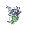
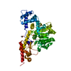
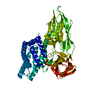
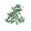
DNA topoisomerase type II (double strand cut, ATP-hydrolyzing) complex / DNA topoisomerase type II (double strand cut, ATP-hydrolyzing) activity / DNA topoisomerase (ATP-hydrolysing) / DNA unwinding involved in DNA replication / DNA topological change / DNA binding / ATP binding / identical protein binding
Similarity search - Function
DNA topoisomerase VI, subunit B / DNA topoisomerase VI, subunit B, transducer / Topoisomerase VI B subunit, transducer / NFACT N-terminal and middle domains / Helicase, Ruva Protein; domain 3 - #50 / Ribosomal Protein S5; domain 2 - #10 / Ribosomal Protein S5; domain 2 / Histidine kinase-like ATPase, C-terminal domain / Heat Shock Protein 90 / Helicase, Ruva Protein; domain 3 ...DNA topoisomerase VI, subunit B / DNA topoisomerase VI, subunit B, transducer / Topoisomerase VI B subunit, transducer / NFACT N-terminal and middle domains / Helicase, Ruva Protein; domain 3 - #50 / Ribosomal Protein S5; domain 2 - #10 / Ribosomal Protein S5; domain 2 / Histidine kinase-like ATPase, C-terminal domain / Heat Shock Protein 90 / Helicase, Ruva Protein; domain 3 / Histidine kinase-, DNA gyrase B-, and HSP90-like ATPase / Histidine kinase-like ATPases / Histidine kinase/HSP90-like ATPase / 2-Layer Sandwich / Orthogonal Bundle / Mainly Alpha / Alpha Beta
Similarity search - Domain/homology
Type 2 DNA topoisomerase 6 subunit B
Similarity search - Component
Biological speciesSulfolobus shibatae (archaea)
MethodX-RAY DIFFRACTION / SYNCHROTRON / MAD / Resolution: 2 Å
AuthorsCorbett, K.D. / Berger, J.M.
CitationJournal: Embo J. / Year: 2003
Title: Structure of the topoisomerase VI-B subunit: implications for type II topoisomerase mechanism and evolution
Authors: Corbett, K.D. / Berger, J.M.
History
DepositionSep 23, 2002Deposition site: RCSB / Processing site: RCSB
Revision 1.0Jan 7, 2003Provider: repository / Type: Initial release
Revision 1.1Oct 16, 2007Group: Version format compliance
Revision 1.2Jul 13, 2011Group: Advisory / Version format compliance
Revision 1.3Feb 14, 2024Group: Data collection / Database references / Derived calculations
Category: chem_comp_atom / chem_comp_bond ...chem_comp_atom / chem_comp_bond / database_2 / pdbx_struct_conn_angle / struct_conn / struct_ref_seq_dif / struct_site
Item: _database_2.pdbx_DOI / _database_2.pdbx_database_accession ..._database_2.pdbx_DOI / _database_2.pdbx_database_accession / _pdbx_struct_conn_angle.ptnr1_auth_seq_id / _pdbx_struct_conn_angle.ptnr3_auth_seq_id / _pdbx_struct_conn_angle.value / _struct_conn.pdbx_dist_value / _struct_conn.ptnr1_auth_comp_id / _struct_conn.ptnr1_auth_seq_id / _struct_conn.ptnr1_label_asym_id / _struct_conn.ptnr1_label_atom_id / _struct_conn.ptnr1_label_comp_id / _struct_conn.ptnr1_label_seq_id / _struct_conn.ptnr2_auth_comp_id / _struct_conn.ptnr2_auth_seq_id / _struct_conn.ptnr2_label_asym_id / _struct_conn.ptnr2_label_atom_id / _struct_conn.ptnr2_label_comp_id / _struct_conn.ptnr2_label_seq_id / _struct_ref_seq_dif.details / _struct_site.pdbx_auth_asym_id / _struct_site.pdbx_auth_comp_id / _struct_site.pdbx_auth_seq_id
Remark 999SEQUENCE The Authors state the residues Y303D and D435N are apparently caused by strain-specific ...SEQUENCE The Authors state the residues Y303D and D435N are apparently caused by strain-specific differences between the strain of S. shibatae and that used for genomic sequencing. The authors state they have cloned the gene several times independently, and the residues are consistently different from the database entry.

-
Structure visualization

Structure viewerMolecule:
MolmilJmol/JSmol

Downloads & links

-
Assembly

Deposited unit
A: Type II DNA topoisomerase VI Subunit B
hetero molecules


Theoretical massNumber of molelcules
Total (without water)53,7193
Polymers53,6381
Non-polymers802
Water4,270237
1


  • Idetical with deposited unit
  • defined by author
TypeNameSymmetry operationNumber
identity operation1_555x,y,z1
Unit cell
Length a, b, c (Å)94.091, 110.968, 54.544
Angle α, β, γ (deg.)90.00, 90.00, 90.00
Int Tables number18
Space group name H-MP21212

-
Components

#1: Protein Type II DNA topoisomerase VI Subunit B / Type II topoisomerase


Mass: 53638.492 Da / Num. of mol.: 1
Source method: isolated from a genetically manipulated source
Source: (gene. exp.) Sulfolobus shibatae (archaea) / Gene: top6b / Production host: Escherichia coli (E. coli) / References: UniProt: O05207, EC: 5.99.1.3
#2: Chemical ChemComp-CA / CALCIUM ION


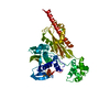
Mass: 40.078 Da / Num. of mol.: 2 / Source method: obtained synthetically / Formula: Ca
#3: Water ChemComp-HOH / water / Water


Mass: 18.015 Da / Num. of mol.: 237 / Source method: isolated from a natural source / Formula: H2O

-
Experimental details

-
Experiment

ExperimentMethod: X-RAY DIFFRACTION / Number of used crystals: 1

-
Sample preparation

CrystalDensity Matthews: 2.65 Å3/Da / Density % sol: 53.63 %
Crystal growTemperature: 293 K / Method: microbatch / pH: 7
Details: PEG 3000, Tris, calcium acetate, pH 7.0, Microbatch, temperature 293K
Crystal grow
*PLUS
Temperature: 20 ℃ / Method: batch method
Components of the solutions
*PLUS
IDConc.Common nameCrystal-IDSol-IDDetailsChemical formula
120 mMTris-HCl11pH7.0
2100 mM11NaCl
312 mg/mlprotein11
420 %PEG300012
5100 mMTris-HCl12pH7.0
6200 mM12Ca(OAc)2

-
Data collection

DiffractionMean temperature: 100 K
Diffraction sourceSource: SYNCHROTRON / Site: ALS / Beamline: 8.3.1 / Wavelength: 1.127 Å
DetectorType: ADSC QUANTUM 4 / Detector: CCD / Date: Dec 20, 2001
RadiationMonochromator: Double crystal / Protocol: SINGLE WAVELENGTH / Monochromatic (M) / Laue (L): M / Scattering type: x-ray
Radiation wavelengthWavelength: 1.127 Å / Relative weight: 1
ReflectionResolution: 2→30 Å / Num. all: 39380 / Num. obs: 37738 / % possible obs: 95.8 % / Observed criterion σ(F): 4 / Observed criterion σ(I): 4 / Rsym value: 0.058 / Net I/σ(I): 16.7
Reflection shellResolution: 2→2.07 Å / Mean I/σ(I) obs: 2.65 / Rsym value: 0.405 / % possible all: 90.6
Reflection
*PLUS
Highest resolution: 2 Å / Lowest resolution: 20 Å / Num. measured all: 284447 / Rmerge(I) obs: 0.058
Reflection shell
*PLUS
Highest resolution: 2 Å / Lowest resolution: 2.07 Å / % possible obs: 90.6 % / Rmerge(I) obs: 0.405 / Mean I/σ(I) obs: 2.7

-
Processing

Software
NameVersionClassification
DENZOdata reduction
SCALEPACKdata scaling
SHARPphasing
REFMAC5refinement
RefinementMethod to determine structure: MAD / Resolution: 2→20 Å / Cor.coef. Fo:Fc: 0.94 / Cor.coef. Fo:Fc free: 0.929 / SU B: 4.541 / SU ML: 0.128 / TLS residual ADP flag: LIKELY RESIDUAL / Cross valid method: THROUGHOUT / σ(F): 0 / ESU R: 0.203 / ESU R Free: 0.167 / Stereochemistry target values: MAXIMUM LIKELIHOOD
RfactorNum. reflection% reflectionSelection details
Rfree0.23809 3130 8.3 %RANDOM
Rwork0.21375 ---
all0.21579 37738 --
obs0.21579 34608 95.97 %-
Solvent computationIon probe radii: 0.8 Å / Shrinkage radii: 0.8 Å / VDW probe radii: 1.4 Å / Solvent model: BABINET MODEL WITH MASK
Displacement parametersBiso mean: 19.944 Å2
Baniso -1Baniso -2Baniso -3
1--1.01 Å20 Å20 Å2
2---1.62 Å20 Å2
3---2.63 Å2
Refinement stepCycle: LAST / Resolution: 2→20 Å
ProteinNucleic acidLigandSolventTotal
Num. atoms3684 0 2 237 3923
Refine LS restraints
Refine-IDTypeDev idealDev ideal targetNumber
X-RAY DIFFRACTIONr_bond_refined_d0.0110.0223762
X-RAY DIFFRACTIONr_bond_other_d
X-RAY DIFFRACTIONr_angle_refined_deg1.3111.9735091
X-RAY DIFFRACTIONr_angle_other_deg
X-RAY DIFFRACTIONr_dihedral_angle_1_deg4.283458
X-RAY DIFFRACTIONr_dihedral_angle_3_deg17.84115737
X-RAY DIFFRACTIONr_chiral_restr0.1070.2567
X-RAY DIFFRACTIONr_gen_planes_refined0.0090.022811
X-RAY DIFFRACTIONr_gen_planes_other
X-RAY DIFFRACTIONr_nbd_refined0.260.31839
X-RAY DIFFRACTIONr_nbd_other
X-RAY DIFFRACTIONr_nbtor_other
X-RAY DIFFRACTIONr_xyhbond_nbd_refined0.1670.5370
X-RAY DIFFRACTIONr_xyhbond_nbd_other
X-RAY DIFFRACTIONr_symmetry_vdw_refined0.3380.358
X-RAY DIFFRACTIONr_symmetry_vdw_other
X-RAY DIFFRACTIONr_symmetry_hbond_refined0.2470.510
X-RAY DIFFRACTIONr_symmetry_hbond_other
X-RAY DIFFRACTIONr_mcbond_it0.7341.52303
X-RAY DIFFRACTIONr_mcangle_it1.4323756
X-RAY DIFFRACTIONr_scbond_it2.55831459
X-RAY DIFFRACTIONr_scangle_it4.4754.51335
X-RAY DIFFRACTIONr_rigid_bond_restr
X-RAY DIFFRACTIONr_sphericity_free
X-RAY DIFFRACTIONr_sphericity_bonded
LS refinement shellResolution: 2→2.051 Å / Total num. of bins used: 20 /
RfactorNum. reflection
Rfree0.304 224
Rwork0.26 2342
Refinement TLS params.

Method: refined / Refine-ID: X-RAY DIFFRACTION

IDL112)L122)L132)L222)L232)L332)S11 (Å °)S12 (Å °)S13 (Å °)S21 (Å °)S22 (Å °)S23 (Å °)S31 (Å °)S32 (Å °)S33 (Å °)T112)T122)T132)T222)T232)T332)Origin x (Å)Origin y (Å)Origin z (Å)
10.4568-0.04050.00730.71450.04750.22230.02630.01540.0041-0.0313-0.0178-0.03090.02220.0286-0.00850.15510.0021-0.00370.1570.00480.134617.473555.579817.3587
25.2598-0.7335-3.07712.56061.26185.1089-0.34960.1221-0.3608-0.13430.1186-0.20130.10390.11370.23090.1689-0.03380.11450.07970.01950.102236.699476.7733-6.4591
30.7291-0.5749-0.28220.71390.31770.2624-0.0058-0.05230.0838-0.02380.017-0.0441-0.0023-0.0079-0.01120.1509-0.014-0.00470.1554-0.00930.155812.92983.524724.5945
Refinement TLS group
IDRefine-IDRefine TLS-IDAuth asym-IDLabel asym-IDAuth seq-IDLabel seq-ID
1X-RAY DIFFRACTION1AA10 - 22911 - 230
2X-RAY DIFFRACTION2AA230 - 310231 - 311
3X-RAY DIFFRACTION3AA311 - 470312 - 471
Refinement
*PLUS
Highest resolution: 2 Å / Lowest resolution: 20 Å / Rfactor Rfree: 0.239 / Rfactor Rwork: 0.214
Solvent computation
*PLUS
Displacement parameters
*PLUS
Refine LS restraints
*PLUS
Refine-IDTypeDev ideal
X-RAY DIFFRACTIONr_bond_d0.011
X-RAY DIFFRACTIONr_angle_d
X-RAY DIFFRACTIONr_angle_deg1.313
LS refinement shell
*PLUS
Highest resolution: 2 Å / Rfactor Rfree: 0.302 / Rfactor Rwork: 0.26

+
About Yorodumi

-
News

-
Feb 9, 2022. New format data for meta-information of EMDB entries

New format data for meta-information of EMDB entries

  • Version 3 of the EMDB header file is now the official format.
  • The previous official version 1.9 will be removed from the archive.

Related info.:EMDB header

External links:wwPDB to switch to version 3 of the EMDB data model

-
Aug 12, 2020. Covid-19 info

Covid-19 info

URL: https://pdbj.org/emnavi/covid19.php

New page: Covid-19 featured information page in EM Navigator.

Related info.:Covid-19 info / Mar 5, 2020. Novel coronavirus structure data

+
Mar 5, 2020. Novel coronavirus structure data

Novel coronavirus structure data

Related info.:Yorodumi Speices / Aug 12, 2020. Covid-19 info

External links:COVID-19 featured content - PDBj / Molecule of the Month (242):Coronavirus Proteases

+
Jan 31, 2019. EMDB accession codes are about to change! (news from PDBe EMDB page)

EMDB accession codes are about to change! (news from PDBe EMDB page)

  • The allocation of 4 digits for EMDB accession codes will soon come to an end. Whilst these codes will remain in use, new EMDB accession codes will include an additional digit and will expand incrementally as the available range of codes is exhausted. The current 4-digit format prefixed with “EMD-” (i.e. EMD-XXXX) will advance to a 5-digit format (i.e. EMD-XXXXX), and so on. It is currently estimated that the 4-digit codes will be depleted around Spring 2019, at which point the 5-digit format will come into force.
  • The EM Navigator/Yorodumi systems omit the EMD- prefix.

Related info.:Q: What is EMD? / ID/Accession-code notation in Yorodumi/EM Navigator

External links:EMDB Accession Codes are Changing Soon! / Contact to PDBj

+
Jul 12, 2017. Major update of PDB

Major update of PDB

  • wwPDB released updated PDB data conforming to the new PDBx/mmCIF dictionary.
  • This is a major update changing the version number from 4 to 5, and with Remediation, in which all the entries are updated.
  • In this update, many items about electron microscopy experimental information are reorganized (e.g. em_software).
  • Now, EM Navigator and Yorodumi are based on the updated data.

External links:wwPDB Remediation / Enriched Model Files Conforming to OneDep Data Standards Now Available in the PDB FTP Archive

-
Yorodumi

Thousand views of thousand structures

  • Yorodumi is a browser for structure data from EMDB, PDB, SASBDB, etc.
  • This page is also the successor to EM Navigator detail page, and also detail information page/front-end page for Omokage search.
  • The word "yorodu" (or yorozu) is an old Japanese word meaning "ten thousand". "mi" (miru) is to see.

Related info.:EMDB / PDB / SASBDB / Comparison of 3 databanks / Yorodumi Search / Aug 31, 2016. New EM Navigator & Yorodumi / Yorodumi Papers / Jmol/JSmol / Function and homology information / Changes in new EM Navigator and Yorodumi

Read more