 Movie
Movie Controller

[English] 日本語
 Yorodumi
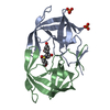
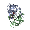
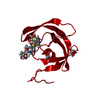
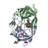
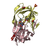
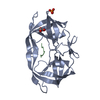
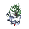
- PDB-1mt9: Viability of a drug-resistant HIV-1 protease mutant: structural i... -
+ Open data

- Basic information
| Entry | Database: PDB / ID: 1mt9 | ||||||
|---|---|---|---|---|---|---|---|
| Title | Viability of a drug-resistant HIV-1 protease mutant: structural insights for better antiviral therapy | ||||||
 Components Components |
| ||||||
 Keywords Keywords | HYDROLASE/HYDROLASE SUBSTRATE / Matrix / Capsid / Gag cleavage / drug resistance / substrate recognition / HYDROLASE / HYDROLASE-HYDROLASE SUBSTRATE COMPLEX | ||||||
| Function / homology |  Function and homology information Function and homology informationHIV-1 retropepsin / symbiont-mediated activation of host apoptosis / retroviral ribonuclease H / exoribonuclease H / exoribonuclease H activity / DNA integration / viral genome integration into host DNA / establishment of integrated proviral latency / RNA-directed DNA polymerase / RNA stem-loop binding ...HIV-1 retropepsin / symbiont-mediated activation of host apoptosis / retroviral ribonuclease H / exoribonuclease H / exoribonuclease H activity / DNA integration / viral genome integration into host DNA / establishment of integrated proviral latency / RNA-directed DNA polymerase / RNA stem-loop binding / viral penetration into host nucleus / host multivesicular body / RNA-directed DNA polymerase activity / RNA-DNA hybrid ribonuclease activity / Transferases; Transferring phosphorus-containing groups; Nucleotidyltransferases / host cell / viral nucleocapsid / DNA recombination / DNA-directed DNA polymerase / aspartic-type endopeptidase activity / Hydrolases; Acting on ester bonds / DNA-directed DNA polymerase activity / symbiont-mediated suppression of host gene expression / viral translational frameshifting / symbiont entry into host cell / lipid binding / host cell nucleus / host cell plasma membrane / virion membrane / structural molecule activity / proteolysis / DNA binding / zinc ion binding Similarity search - Function | ||||||
| Biological species |   Human immunodeficiency virus 1 Human immunodeficiency virus 1 | ||||||
| Method |  X-RAY DIFFRACTION / FOURIER SYNTHESIS / Resolution: 2 Å X-RAY DIFFRACTION / FOURIER SYNTHESIS / Resolution: 2 Å | ||||||
 Authors Authors | Prabu-Jeyabalan, M. / Nalivaika, E.A. / King, N.M. / Schiffer, C.A. | ||||||
 Citation Citation | Journal: J.Virol. / Year: 2003 Title: Viability of drug-resistant human immunodeficiency virus type 1 protease variant: structural insights for better antiviral therapy Authors: Prabu-Jeyabalan, M. / Nalivaika, E.A. / King, N.M. / Schiffer, C.A. #1: Journal: J.Mol.Biol. / Year: 2000Title: How does a symmetric dimer recognize an asymmetric substrate? A substrate complex of HIV-1 protease Authors: Prabu-Jeyabalan, M. / Nalivaika, E.A. / Schiffer, C.A. #2: Journal: Structure / Year: 2002Title: Substrate shape determines specificity of recognition for HIV-1 protease: Analysis of crystal structures of six substrate complexes Authors: Prabu-Jeyabalan, M. / Nalivaika, E.A. / Schiffer, C.A. #3: Journal: Protein Sci. / Year: 2002Title: Lack of synergy for inhibitors targeting a multi-drug resistant HIV-1 protease Authors: King, N.M. / Melnick, L. / Prabu-Jeyabalan, M. / Nalivaika, E.A. / Yang, S.-S. / Gao, Y. / Nie, X. / Zepp, C. / Heefner, D.L. / Schiffer, C.A. | ||||||
| History |
|
- Structure visualization
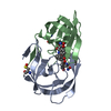
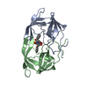
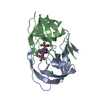
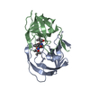
| Structure viewer | Molecule: MolmilJmol/JSmol |
|---|
- Downloads & links
-Download
| PDBx/mmCIF format | 1mt9.cif.gz | 55.2 KB | Display | PDBx/mmCIF format |
|---|---|---|---|---|
| PDB format | pdb1mt9.ent.gz | 39.7 KB | Display | PDB format |
| PDBx/mmJSON format | 1mt9.json.gz | Tree view | PDBx/mmJSON format | |
| Others |  Other downloads Other downloads |
-Validation report
| Arichive directory | https://data.pdbj.org/pub/pdb/validation_reports/mt/1mt9ftp://data.pdbj.org/pub/pdb/validation_reports/mt/1mt9 | HTTPS FTP |
|---|
-Related structure data
| Related structure data | 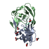 1mt7C  1mt8C 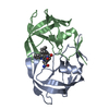 1mtbC 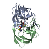 1n49C  1mtrS S: Starting model for refinement C: citing same article ( |
|---|---|
| Similar structure data |

-Links
 PDBj
PDBj

- Assembly
| Deposited unit | 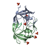
| ||||||||||
|---|---|---|---|---|---|---|---|---|---|---|---|
| 1 |
| ||||||||||
| Unit cell |
| ||||||||||
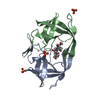
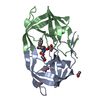
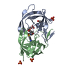
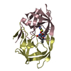
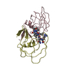
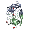
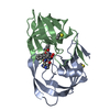
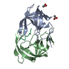
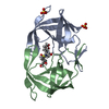
| Details | The entire dimer is provided in the coordinate set and the monomers are distinguished by the chain IDs `A' and `B'. The decamer sequence corresponding to the MA-CA cleavage site of HIV-1 Gag polyprotein. |
-Components
| #1: Protein | Mass: 10772.724 Da / Num. of mol.: 2 / Mutation: Q7K, D25N, L63P, V82A Source method: isolated from a genetically manipulated source Source: (gene. exp.) Human immunodeficiency virus 1 / Genus: Lentivirus / Plasmid: pXC35-pEN18 / Production host:  #2: Protein/peptide | | Mass: 1173.325 Da / Num. of mol.: 1 / Source method: obtained synthetically Details: This peptide sequence occurs naturally in Gag of HIV-1 #3: Chemical | ChemComp-PO4 /   Mass: 94.971 Da / Num. of mol.: 6 / Source method: obtained synthetically / Formula: PO4 Mass: 94.971 Da / Num. of mol.: 6 / Source method: obtained synthetically / Formula: PO4#4: Water | ChemComp-HOH / |  Mass: 18.015 Da / Num. of mol.: 88 / Source method: isolated from a natural source / Formula: H2O Mass: 18.015 Da / Num. of mol.: 88 / Source method: isolated from a natural source / Formula: H2O |
|---|
-Experimental details
-Experiment
| Experiment | Method: X-RAY DIFFRACTION / Number of used crystals: 1 |
|---|
- Sample preparation
| Crystal | Density Matthews: 2.06 Å3/Da / Density % sol: 40.41 % | ||||||||||||||||||||||||||||||
|---|---|---|---|---|---|---|---|---|---|---|---|---|---|---|---|---|---|---|---|---|---|---|---|---|---|---|---|---|---|---|---|
| Crystal grow | Temperature: 298 K / Method: vapor diffusion, hanging drop / pH: 6.2 Details: sodium phosphate, sodium citrate, ammonium sulphate, pH 6.2, VAPOR DIFFUSION, HANGING DROP at 298K | ||||||||||||||||||||||||||||||
| Crystal grow | *PLUS | ||||||||||||||||||||||||||||||
| Components of the solutions | *PLUS
|
-Data collection
| Diffraction | Mean temperature: 298 K |
|---|---|
| Diffraction source | Source: ROTATING ANODE / Type: RIGAKU / Wavelength: 1.5418 Å |
| Detector | Type: RIGAKU RAXIS IV / Detector: IMAGE PLATE / Date: Nov 30, 1999 / Details: Yale mirrors |
| Radiation | Monochromator: Yale Mirrors / Protocol: SINGLE WAVELENGTH / Monochromatic (M) / Laue (L): M / Scattering type: x-ray |
| Radiation wavelength | Wavelength: 1.5418 Å / Relative weight: 1 |
| Reflection | Resolution: 2→32.84 Å / Num. all: 12659 / Num. obs: 12659 / % possible obs: 95.4 % / Observed criterion σ(F): 0 / Observed criterion σ(I): 0 / Biso Wilson estimate: 12.4 Å2 / Rmerge(I) obs: 0.09 / Net I/σ(I): 7.7 |
| Reflection shell | Resolution: 2→2.07 Å / Rmerge(I) obs: 0.33 / Num. unique all: 1182 / % possible all: 92 |
| Reflection | *PLUS Highest resolution: 2 Å / Num. measured all: 98506 / Rmerge(I) obs: 0.09 |
| Reflection shell | *PLUS Highest resolution: 2 Å / % possible obs: 92 % / Num. unique obs: 1182 |
- Processing
| Software |
| ||||||||||||||||||||||||||||||||||||||||||||||||||||||||||||
|---|---|---|---|---|---|---|---|---|---|---|---|---|---|---|---|---|---|---|---|---|---|---|---|---|---|---|---|---|---|---|---|---|---|---|---|---|---|---|---|---|---|---|---|---|---|---|---|---|---|---|---|---|---|---|---|---|---|---|---|---|---|
| Refinement | Method to determine structure: FOURIER SYNTHESIS Starting model: 1MTR Resolution: 2→32.84 Å / Rfactor Rfree error: 0.006 / Data cutoff high absF: 172172.14 / Data cutoff low absF: 0 / Isotropic thermal model: RESTRAINED / Cross valid method: THROUGHOUT / σ(F): 0 / σ(I): 0 / Stereochemistry target values: Engh & Huber
| ||||||||||||||||||||||||||||||||||||||||||||||||||||||||||||
| Solvent computation | Solvent model: FLAT MODEL / Bsol: 67.3885 Å2 / ksol: 0.367432 e/Å3 | ||||||||||||||||||||||||||||||||||||||||||||||||||||||||||||
| Displacement parameters | Biso mean: 23.8 Å2
| ||||||||||||||||||||||||||||||||||||||||||||||||||||||||||||
| Refine analyze | Luzzati coordinate error free: 0.26 Å / Luzzati sigma a free: 0.18 Å | ||||||||||||||||||||||||||||||||||||||||||||||||||||||||||||
| Refinement step | Cycle: LAST / Resolution: 2→32.84 Å
| ||||||||||||||||||||||||||||||||||||||||||||||||||||||||||||
| Refine LS restraints |
| ||||||||||||||||||||||||||||||||||||||||||||||||||||||||||||
| LS refinement shell | Resolution: 2→2.07 Å / Rfactor Rfree error: 0.028 / Total num. of bins used: 10
| ||||||||||||||||||||||||||||||||||||||||||||||||||||||||||||
| Xplor file |
| ||||||||||||||||||||||||||||||||||||||||||||||||||||||||||||
| Refinement | *PLUS Highest resolution: 2 Å / Rfactor Rfree: 0.21 | ||||||||||||||||||||||||||||||||||||||||||||||||||||||||||||
| Solvent computation | *PLUS | ||||||||||||||||||||||||||||||||||||||||||||||||||||||||||||
| Displacement parameters | *PLUS | ||||||||||||||||||||||||||||||||||||||||||||||||||||||||||||
| Refine LS restraints | *PLUS
| ||||||||||||||||||||||||||||||||||||||||||||||||||||||||||||
| LS refinement shell | *PLUS Highest resolution: 2 Å |
